Indication
Assessment of the airway
Suspicion of a foreign body
Malformation of the upper airway
Obstructive sleep apnea syndrome
Persistent/recurrent symptom (cough, stridor, wheezing)
Persistent/recurrent disease (atelectasis, pneumonia, etc.)
Follow-up of some conditions (tracheostomy, transplantation, surgery)
Bronchoalveolar lavage
Pulmonary hemorrhage
Interstitial lung disease
Pneumonia in immunosuppressed patients
Severe pneumonia in immunocompetent patients
Suspicion of endobronchial tuberculosis
Suspicion of pulmonary aspiration
Therapeutic indications for flexible bronchoscopy
Indication |
|---|
Aspiration of a foreign body |
Assessment before and after extraction of a foreign body Extraction of a distal foreign body |
Bronchoalveolar lavage |
Expansion of persistent or massive atelectasis Inhalation of highly irritating matter (barium, soot, blood, etc.) Selective/massive cleaning of alveolar proteinosis Application of medication (DNase, surfactant, cold serum, etc.) |
Difficult intubation |
Monointubation for thoracic surgery Seldinger endoscopic intubation technique |
Additional procedures (unusual indications) |
Airway biopsy Distal airway laser surgery Dilatation of the airway with an inflatable balloon Sealing of a persistent tracheoesophageal fistula |
Contraindications
The contraindications for flexible bronchoscopy are generally relative, because these situations will be transitory; therefore, they do not involve major risk. The only real contraindications are scenarios in which the procedure will not be useful diagnostically or the patient’s guardians do not give consent for its use.
- 1.
Hemodynamic instability
- 2.
Intracranial hypertension
- 3.
Hemorrhagic diathesis
- 4.
Pulmonary hypertension
- 5.
Massive hemoptysis
- 6.
Respiratory insufficiency
Technical Aspects
Equipment
There are two types of instruments currently available: the fiber bronchoscope, which has an optical fiber that sends images to a simple viewer or an external camera and then to an image processor; and the more recently introduced video endoscope, featuring a camera at one end of the instrument that sends images directly to a processor. The latter instrument has better optical resolution, being termed “high definition”, when the image is translated into a determined number of pixels on a screen. Both types of equipment should include a working channel for aspiration of secretions, instrumentalization, and drug instillation.
Types and sizes of instruments, their applicability in different age groups, and their compatibility with other equipment
Patient age | Instrument type | External diameter (mm) | Working Channel (mm) | BAL | Biopsy | Laryngeal mask (minimum size (mm)) | Endotracheal tube (minimum size (mm)) |
|---|---|---|---|---|---|---|---|
Premature/newborn | Fiberscope | 2.2 | No | No | No | 1 | ≥3 |
0–2 years | Fiberscope/video endoscope | 2.7–2.8 | 1.2 | Yes | Yesa | 1.5 | ≥3.5 |
0–5 years | Fiberscope/video endoscope | 3.4–3.6 | 1.2 | Yes | Yesa | 2 | ≥4.5 |
2–5 years | Fiberscope | 4 | 2 | Yes | Yes | 2.5 | ≥5.5 |
>5 years | Fiberscope/video endoscope | 4.9–5.1 | 2–2.2 | Yes | Yes | 3 | ≥6 |
>15 years | Fiberscope/video endoscope | 5.9–6 | 2.2–2.8 | Yes | Yes | 4 | ≥7.5 |
Procedure
Flexible bronchoscopy is a clean procedure, but it is not sterile, because the route of entry—nasal, tracheostomy, laryngeal mask, etc.—is always contaminated. The risk of intrahospital infection by flexible bronchoscopy is low if protocols for disinfection, storage, and handling of equipment are adequately followed. Sterilization can be done with ethylene oxide, ortho-phthalaldehyde, or glutaraldehyde for high-level disinfection. In this scenario, disinfection with glutaraldehyde is very practical, given that it takes only 60 minutes to have the equipment available for reuse, but it does require a central unit with staff trained to ensure adherence to standards of efficacy, as well as the safety of the personnel who handle the disinfectant. There is now equipment that includes a disposable outer jacket with a suction channel. The external jacket is sterile and is discarded after the procedure; thus, multiple procedures can be conducted without the need for disinfection intervals.
First, the procedure is explained in detail to the parents before it begins, and informed consent is obtained in accordance with the protocols of the specific center, with the objective of clarifying the procedure, its usefulness, and potential complications. The procedure room should be equipped with oxygen, central aspiration, a resuscitation cart, a manual ventilator, a laryngoscope, tracheal tubes, masks, aspiration cannulas, monitors, drugs for sedation, and inputs. Other situations in which flexible bronchoscopy is applied are the critical care unit and the operating room, which allows us to carry out the procedure without any additional risk. The procedure is carried out with noninvasive cardiovascular monitoring (heartbeat and blood pressure) and continuous registering of peripheral oxygen saturation (SpO2) until the patient recovers completely. The procedure should be carried out by a doctor trained in endoscopy, accompanied by another doctor trained in deep sedation and monitoring. It is also necessary to have the help of a nurse and a high-level technician trained in the procedure, administration of drugs, and high-level disinfection. The members of the team have specific responsibilities during the procedure, which are coordinated prior to the procedure, such as drug labeling, registration of vital signs, and positioning of the patient, nasal or oral aspiration of secretions, and assisting the operator in other tasks.
Sedation
The patient should have a clinical assessment for potential problems associated with the procedure, including a consultation between an anesthesiologist and the doctor in charge of sedation for the procedure. The patient should have a permeable venous access inserted before the procedure begins. The sedation depends on the access and the characteristics of the patient, and can range from deep sedation with access through the nose (with spontaneous ventilation) to profound anesthesia and a muscular block (with assisted manual ventilation with the airway maintained). For patients at risk of respiratory insufficiency or a minimal ventilatory reserve, the use of a laryngeal mask, an endotracheal tube, or manual ventilation should be considered before the procedure is conducted. A laryngeal mask can be used in procedures in which the objective is not to view the airway dynamics but, rather, to perform more invasive exploration, whether it is to search for a foreign body or to perform BAL for culturing, bronchial brushing, or bronchial biopsy. The idea is to anticipate hypoxemia to avoid systemic consequences. The most useful medications for this sedation are those that, in combination, offer sedation and analgesia for short periods, with minimal effects on spontaneous respiration. The most widely used medications are midazolam with morphine, midazolam with ketamine, and propofol. To some degree, and depending on their doses, all of the medications that are used can have a depressive effect on the respiratory system and increase secretions in the airway; thus, there are adverse effects that must be monitored throughout the procedure. Topical sedation is necessary from the nasal passage to the main carina to avoid pain, reflexive spasms, and coughing. Lidocaine in either a liquid or gel form is used to an obtain an adequate level of sedation. Lidocaine is preferably administered at 2% in the upper airway and 1% in the lower airway, and not exceeding a total dose of 7 mg/kg. The administration of lidocaine in the upper airway should be preceded by adequate visualization of the epiglottis and of the movement of the vocal cords, so as to not attribute exaggerated collapse of these structures to a pathological condition rather than to the effect of anesthesia, especially in patients in whom paralysis of the vocal cords is suspected.
Administration of atropine, which was a common practice in instrumentation of the airway in the past, is now reserved for patients who present an excessive vagal response or for infant under 6 months of age. Administration of oxygen is considered in all procedures from the beginning because of the high risk of hypoxemia when the instrument is inserted into the airway, to which is added an exaggerated spasm reflex, coughing, and the excess of secretions produced by the introduction of the instruments. Oxygen is provided by means of a nasal cannula with a flow of ≥2 l/min, so as to obtain a reservoir of the inhaled fraction of oxygen in the rhinopharyngeal space of a patient breathing spontaneously.
Another way to administer oxygen is by manual ventilation with an inflatable bag, whether through an endotracheal tube or a laryngeal mask. Finally, oxygen can be administered for bronchoscopy. However, this should be done only in the central airway and intermittently, because of the high risk of increasing pressure in the airway and an associated pneumothorax. Ideally, an SpO2 of >93% is maintained continuously and the procedure is temporarily interrupted if this level is not maintained. In this context, hypoxemia is always considered a late indicator of hypoventilation because oxygen is administered concomitantly throughout the procedure.
Bronchoalveolar Lavage
Bronchoalveolar lavage consists of administration of a physiological serum in a specific area for diagnostic or therapeutic purposes. The procedure involves positioning the point of the instrument in a bronchus with a caliber similar to that of the device, such that it allows for a certain degree of bronchial sealing when the physiological serum is transferred, which is termed “interlocking.” The procedure is done in the selected area either because it is a collapsed pulmonary area (in the case of lobar atelectasis) or because it presents infectious compromise, in which case the procedure is expected to determine the etiological agent (pneumonia in the case of immunocompromised patients). Upon reaching the desired location, 1–2 ml/kg of physiological serum is instilled in the working channel on at least three occasions (with a volume not greater than 25% of the functional residual capacity), to be subsequently aspirated through the same channel, with recovery of approximately 30–50% of the instilled volume. If a good seal is achieved during the maneuver, the liquid that is obtained contains cells from the respiratory bronchiole and the alveolar territory.
The risk of contamination of equipment that enters the upper airway can be reduced with the use of a laryngeal mask and with bronchial brushing, using brushes with protected ends that are only exposed when they reach the area to obtain a culture sample.
Recovery
The patient recovers from flexible bronchoscopy in the procedure room, the critical care unit, or the anesthetic recovery room, under the same noninvasive monitoring that is described above, and under the responsibility of the physician in charge of sedation. Once the patient awakens from sedation and his or her respiratory and cardiovascular condition is stable, he or she is taken to the unit of origin. Depending on the sedation that is used, and once the patient is fully awake, he or she can be fed within 2 hours after the procedure. This safety interval also allows for full recovery of the protective reflexes of the airway following the use of local lidocaine. A routine chest x-ray is not necessary unless there is respiratory deterioration with increased oxygen requirements and breathing difficulty. Following the application of flexible bronchoscopy with BAL, the patient can present a fever, which is usually limited and is not associated with any intercurrent infection. This can be prevented with administration of paracetamol (15 mg/kg every 6–8 hours, orally) within hours after the procedure or dexamethasone (0.3 mg/kg) during the procedure.
Complications of Flexible Bronchoscopy
Complications of flexible bronchoscopy are common; most of them are transitory. The complications are directly related to the duration of the procedure, because of which it is vital to reduce the procedural time as much as possible. The most common complications are the result of the partial occlusion of the airway and the effects of sedation. The most frequent complication is transitory hypoxemia, which occurs as a result of hypoventilation while the equipment is in the airway, and it is resolved by temporarily removing the equipment. An excessive cough reflex is also common, which is generally controlled with the application of local lidocaine. The use of topical anesthesia in patients with excessive secretions (e.g., due to cystic fibrosis) is decisive to prevent this complication. Less common, but no less important, are complications arising from the sedation, such as hypoventilation secondary to the use of depressor drugs and accumulation of secretions due to the lack of a cough reflex. Rapid diagnosis allows for immediate resolution with regard to the positioning of the upper airway, manual ventilation with a mask, and sometimes intubation of the airway. In patients with respiratory difficulty prior to the procedure, endotracheal intubation and mechanical ventilation should be considered. Epistaxis during or after the application of flexible bronchoscopy is common in infants, where the narrow nasal passage is similar in size to the equipment. This condition often resolves spontaneously or after application of cold serum at the end of the procedure. Other complications are also common, such as postprocedural hemorrhaging of the distal airway, pneumothorax, laryngospasms, and pneumonia.
Performance of Flexible Bronchoscopy
Congenital Stridor
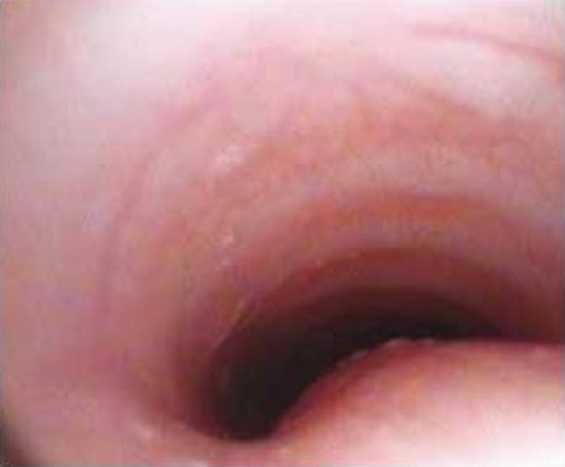
Most patients who consult physicians because of a congenital stridor have laryngomalacia, a condition that is suspected on the basis of a characteristic medical history, which can be confirmed by nasal fibroscopy conducted with topical anesthesia. However, flexible bronchoscopy is necessary to confirm the causes of the condition in patients with a severe stridor, episodes of cyanosis or apnea, or with eating difficulties. Likewise, flexible bronchoscopy should be applied when the stridor has an uncommon temporal profile, when a condition other than laryngomalacia is suspected, and, above all, when there is a therapeutic possibility. The procedure is sometimes justified by parental anxiety caused by the stridor. Common causes are vocal cord paralysis and subglottic stenosis, both of which are generally related to a previous treatment: surgery for patent ductus arteriosus in the former case, and prolonged intubation in the latter case. More uncommon conditions that are nevertheless important to rule out are a tracheal cleft, laryngeal membrane, tracheal stenosis, and tracheal hemangioma.
Acute Stridor
In patients suffering from an episode of acute obstructive laryngitis, flexible bronchoscopy is justified only when the patient has not improved despite treatment or when intubation is needed because of deterioration of the condition. The findings can be compatible with bacterial tracheitis, a foreign body, or unexpected congenital malformations, which is particularly relevant information for treating the patient.
Recurrent Laryngitis
If there are more than three recurrent episodes of acute obstructive laryngitis, especially when they occur within a year, a flexible bronchoscopy study is mandatory. There is evidence from such cases in children under 3 years of age that a third of them present some degree of alteration of the airway that explains the recurrence of this condition.
Upper Airway Obstruction
Assessment of the upper airway of patients with upper airway obstruction (UAO) is facilitated by directed viewing through a flexible bronchoscopy, where the potential therapeutic value of medical or surgical solutions in specific situations can be evaluated. In patients with craniofacial malformations or neuromuscular disease, repositioning of the tongue or collapse of the tonsils can be very evident with the patient under sedation but not when the patient is awake. The procedure in this scenario tends to be highly risky because of the possibility of destabilizing the airway.
Atelectasis
In the context of lobar or massive atelectasis, flexible bronchoscopy has a mainly diagnostic objective, whether this situation is the result of a bronchial malformation, vascular compression, a foreign body, a mucosal plug, or an endobronchial lesion. In conditions in which bronchial cleaning with physiological serum facilitates the expansion of the tributary area, bronchial lavage, which expands the area in 70% of cases, can be attempted. It is useful to apply diluted adrenaline (epinephrine) and/or DNase in this procedure to facilitate the removal of mucus plugs. In other situations, the treatment is done according to the endoscopic findings.
Pneumonia in Immunocompetent Patients
The performance of BAL through a flexible bronchoscope in patients with an acute lower respiratory infection is variable but does not achieve etiological identification in more than 50% of cases. The performance is affected by prior use of antibiotics, the timing of the disease, and the quality of the samples obtained. New techniques such as polymerase chain reaction and quantitative bacterial culturing have improved etiological identification. The procedure is thus justified in patients who have a severe condition without a known agent and who do not respond to treatment.
Pneumonia in Immunocompromised Patients
In immunocompromised patients, pulmonary infections by the usual and opportunistic agents are a common cause of complications, and their identification is usually difficult. The agents involved vary according to when the disease appears and its treatment: neutropenia, chemotherapy, antibiotics, etc. BAL is effective in more than 50% of cases under these conditions, and it is even more justified in immunocompromised patients because noninvasive tests have poorer results. Bronchial lavage has a high negative predictive value for some infectious agents, so therapeutic decisions can be facilitated by reducing antibiotic coverage. However, the time available to perform the procedure may be limited by the presence of hypoxemic respiratory failure because of the high risk, under these conditions, of the patient’s condition worsening and the need to resort to ventilatory support. Other complications are more common in immunocompromised patients; thus, the potential benefits must always be weighed against the risks.
Recurrent Wheezing and Pneumonia
Stay updated, free articles. Join our Telegram channel

Full access? Get Clinical Tree


