Obturator Foramen Bypass Grafts in Groin Sepsis


Obturator Foramen Bypass Grafts in Groin Sepsis


Bruce A. Perler


In an era of rapid evolution and dramatic innovation in the diagnosis and management of vascular disease, infection remains one of the most serious life- and limb-threatening problems for the patient and difficult challenges for the surgeon. Although a variety of options have been used to treat vascular graft infection, including in situ replacement with autogenous conduits, antibiotic-impregnated synthetic grafts, cryopreserved grafts, homografts, and rotational muscle flaps, the conventional approach remains resection of the infected graft and surrounding tissues with an extra-anatomic bypass. The majority of vascular infections manifest in the groin, and the extra-anatomic obturator bypass continues to be a valuable strategy for treating patients in this setting.


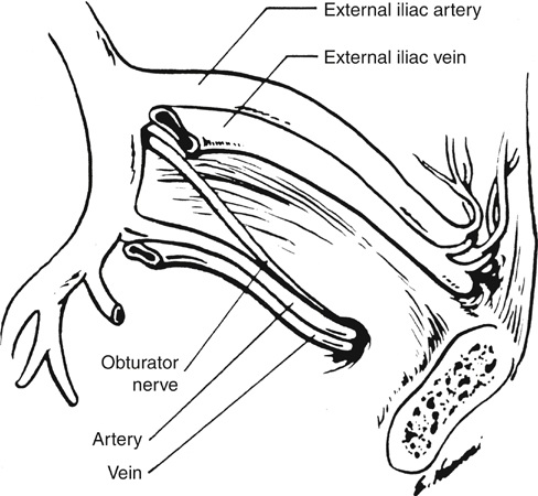
Routing of an arterial bypass graft from the retroperitoneum into the leg through the obturator foramen was introduced by Shaw and Baue in 1963. Although the technical details of the procedure have been modified over the last nearly 5 decades, its fundamental rationale has remained the same, namely, to provide inflow to the extremity while excluding the new graft from communication with the femoral vessels in the groin. Similarly, although the indications for obturator foramen bypass have broadened somewhat, the procedure is most often used to manage groin sepsis caused by an infected previous graft, an infected false or true femoral aneurysm, or some other suppurative process (Box 1).


Although conventional bypass grafts are being increasingly supplanted by endovascular procedures, groin sepsis will continue to be encountered in practice. For example, infection is now being reported as an uncommon complication in association with arterial closure devices. Therefore, obturator foramen bypass will remain an important and potentially life- and limb-salvaging option to treat vascular sepsis.


Patient Selection


A fundamental prerequisite for a successful outcome of obturator bypass in the patient with arterial infection in the groin is that the septic process be truly confined to the groin. This approach is most appropriate for the patient with native femoral artery infection. Although rarely a patient has an infected arteriosclerotic femoral artery aneurysm or septic arteritis resulting from previous vascular cannulation for a diagnostic arteriogram or an endovascular procedure, a more common scenario is the presence of a mycotic pseudoaneurysm in an illicit drug user.


Obturator foramen bypass is also a viable option in the patient with infection involving a previously placed prosthetic bypass graft. For example, it is an excellent procedure for providing inflow to the recipient limb in the patient who requires excision of an infected femorofemoral bypass graft. In this situation the native iliac artery (ipsilateral or contralateral) or aorta should be uninvolved with the infectious process. On the other hand, one must be cautious in planning an obturator bypass in the patient with infection involving the patent limb of an aortofemoral bypass because infection occurring in the groin in this situation often tracks proximally along the entire extent of that graft limb, and in fact the main body of the graft may be involved. In this situation the proximal extent of the septic process precludes performing the proximal anastomosis of the new graft in a clean field. Similarly, when the patient has an infection involving the proximal segment of an infrainguinal bypass graft in the groin, it is important to rule out extension of the septic process along the more distal aspect of the conduit so that one can perform the distal anastomosis of the obturator bypass at an infection-free site.


The acuteness of the clinical presentation influences the completeness of the preoperative workup. In the stable patient with graft infection in the groin, the author obtains a computed tomography (CT) scan to assess proximal or distal extension of the septic process. The presence of perigraft gas or fluid and loss of the perigraft fat signal are presumptive evidence of infection. Alternatively, magnetic resonance imaging (MRI) can provide similar evidence of perigraft infection. The CT or MRI should be followed by arteriography both to identify any unsuspected anastomotic aneurysms proximal or distal to the groin and to assess the infrainguinal vascular anatomy to select the most appropriate site for the distal anastomosis of the obturator foramen bypass graft.


Operative Procedure: Anatomic Considerations


The patient is placed in the supine position with the operative side elevated approximately 30 degrees on folded sheets under the ipsilateral buttock; the hip and leg are abducted and externally rotated. A Foley catheter is placed to decompress the bladder. In the absence of frank femoral hemorrhage, the obturator bypass should be constructed initially, and then the infected graft is removed or the infected femoral artery is repaired secondarily through a separate groin incision. Thus the involved groin must be completely isolated from the obturator foramen bypass operative field with adherent plastic drapes and covered with a sterile towel secured to the underlying skin. The operative field is then prepared with an antiseptic solution and draped to include the entire abdomen and both lower extremities.


Exploration of the proximal limb of an aortofemoral graft (or the iliac arteries) on the involved side may be carried out through a midline or paramedian transperitoneal incision or a curvilinear flank incision. The author prefers the latter, muscle-splitting approach and exploration of the intended site of the proximal anastomosis through the retroperitoneum.


Safe construction of an obturator bypass requires familiarity with anatomic planes not often encountered by the vascular surgeon. In brief, the obturator nerve and the obturator artery and vein pass through the superior and lateral aspect of the obturator membrane and are the key landmarks in identifying the foramen. Although the obturator artery arises from the anterior division of the internal iliac artery in most people (Figure 1), in nearly 30% of patients it arises entirely, or in part, from the inferior epigastric artery. In this case, the vessel courses posteriorly over the superior ramus of the pubis, often accompanied by a small vein, and is vulnerable to injury if not carefully identified. The prominent obturator internus muscle overlies the inferior aspect of the foramen.


Stay updated, free articles. Join our Telegram channel

Aug 25, 2016 | Posted by in CARDIOLOGY | Comments Off on Obturator Foramen Bypass Grafts in Groin Sepsis

Full access? Get Clinical Tree

Get Clinical Tree app for offline access