Obesity Hypoventilation Syndrome

Chapter 62 Obesity Hypoventilation Syndrome


Obesity hypoventilation syndrome is a disorder characterized by the presence of daytime compensated respiratory acidosis (i.e., hypercapnia with normal pH and increased serum bicarbonate), in patients in whom no cause of ventilatory failure can be identified. These patients cannot ensure a normal minute ventilation even in the resting state, although clinical evaluation will rule out disorders associated with decreased muscle force (neuromuscular diseases), abnormal muscle geometry (e.g., kyphoscoliosis), severe airway obstruction and abnormal ventilation-perfusion relationships (e.g., chronic obstructive pulmonary disease [COPD], emphysema), or parenchymal abnormalities (e.g., interstitial lung diseases). In these patients, with performance of a respiratory maneuver consisting of serial deep inspirations, subsequently the arterial carbon dioxide tension (PaCO2) normalizes, and in those in whom hypoxia is present, oxygen saturation (SaO2) rises by 4% to 6% in less than 2 minutes. Of note, however, they do not sustain adequate minute ventilation and remain hypercapnic, with the kidneys compensating to maintain a normal pH. Another clinical element quite evident is that such persons are obese, frequently morbidly obese, with body mass index (BMI) values in excess of 35 kg/m2. It is the association of pathologic obesity and daytime hypercapnia that defines the obesity hypoventilation syndrome.


When these patients are studied in a sleep laboratory, it becomes clear that they are afflicted with some form of sleep-disordered breathing pattern. The vast majority of patients have a moderate to severe form of obstructive sleep apnea (OSA), with a minority showing no obstructed breathing but simply sleep-related hypoxia and hypercapnia (long periods of SaO2 below 90% and an increase in PaCO2 of about 10 mm Hg developing overnight). The diagnosis of obesity hypoventilation syndrome can be reached with certainty only when other causes of daytime hypercapnia have been excluded.


Risk Factors


Several conditions seem to predispose an obese person to development of the obesity hypoventilation syndrome. Undisputedly, in comparison with patients with OSA, patients diagnosed with this syndrome seem to be more obese, and they have more severe OSA. The main cause of obesity is well recognized to be an excess of food intake in relation to the energy expenditure requirements of the organism, resulting in the constitution of an energy stock essentially in the form of fat deposits. According to worldwide epidemiologic data, the prevalence of obesity is increasing everywhere, and this trend seems to be related essentially to a rapid change in eating habits, themselves influenced by technologic changes and food industry policies. Whether this reflects an improvement in the standard of living is a matter of debate.


The health consequences of obesity should prompt implementation of educational strategies to promote adequate behavioral changes in eating habits. Nevertheless, at the individual level, genetic factors may have an influence on the development of obesity, depending on the balance between what can be termed “catabolic” and “anabolic” genes. More than 250 genes have been identified that have a more or less important influence on the final handling of the energetic balance. The clinical importance of each genetic variant, however, is not yet well appreciated. Only a few monogenetic causes of obesity have been described in humans, the best example being mutations in the gene coding for the melanocortin 4 receptor. It has to be stressed, however, that obesity will not develop if access to food is compromised, or if energy expenditure exceeds energy absorption.


An apnea-hypopnea index (AHI) greater than 50, as measured by the number of apneic or hypopneic episodes per hour of sleep, also seems to be a risk factor for obesity hypoventilation syndrome, because 25% of patients with such AHIs in several studies had the syndrome. Oxygen saturation nadir levels below 60% during polysomnography and moderate to severe restriction found on pulmonary function testing also are predictors for the syndrome, with odds ratios of 4 and 10, respectively. Finally, some studies found that patients with obesity hypoventilation syndrome had greater neck, waist, and hip circumferences and a larger waist-to-hip ratio than their counterparts with OSA.


Pathophysiology


Obese patients, both eucapnic and hypercapnic, have significant reductions in functional residual capacity and expiratory reserve volume with preservation of inspiratory capacity and often normal or slightly reduced total lung capacity. But obesity is not, however, the only determinant of hypoventilation, because only a third of morbidly obese persons develop hypercapnia. Some differences between eucapnic obese and hypercapnic subjects have been recognized and are summarized next.


Obstructive Sleep Apnea


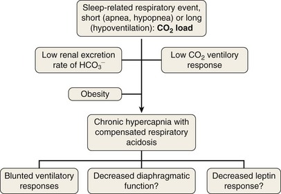
OSA may play a role in the pathogenesis of chronic hypercapnia in those patients, because in most cases, treatment of OSA corrects the hypercapnia. Indeed, to compensate for the effect of intermittent periodic breathing and resultant acute hypercapnia, normal subjects as well as patients with OSA will increase their tidal volume in the first breath after an apnea (hyperventilation). With physiologic differences in ventilatory responses between subjects, however, an overload in CO2 could result. The duration of the apneas also could contribute: When apneic episode duration becomes three times longer than the breathing interval, CO2 tends to accumulate despite maximal tidal volume, because there is insufficient time for adequate hyperventilation events (Figure 62-1). In addition, hypercapnia could blunt the ventilatory responses: The initial ventilation after an apneic episode is directly related to the volume of CO2 loaded during the preceding respiratory event and thus represents an index of CO2 ventilatory response. Hypercapnic patients demonstrated depression of this index of ventilatory compensation compared with that in eucapnic patients. It has been shown that the apnea-to-interapnea duration ratio is greater in hypercapnic patients than in eucapnic patients.


One study also has shown impaired CO2 homeostasis after respiratory events may be mediated by opioids or opioid receptors, because endorphin blockade changed this pattern. Increased cerebrospinal fluid beta-endorphin activity with return to normal values has been reported in subjects with OSA. This finding could explain hypercapnia on awakening after a night full of apneic episodes. To understand why in a period of wakefulness free of apnea the PaCO2 does not normalize, the role of the renal system has to be considered. Hypercapnia leads to respiratory acidosis, which activates the process of renal compensation.


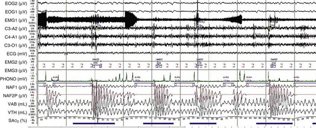
To elucidate the mechanisms that are involved in the development of hypercapnia in patients with OSA, Norman and co-workers have proposed, using a computer model, some hints for prediction of the transition from acute hypercapnia during sleep-disordered breathing (apnea, hypopnea, and hypoventilation) to chronic daytime hypercapnia. In their model, when the ventilatory CO2 response and renal HCO3 excretion were normal, increases in PaCO2 and HCO3 did not develop. The bicarbonate excretion during the day compensated for that retained during the night. When CO2 ventilatory response was very low, however, the model demonstrated a modest rise in PaCO2 and HCO3 measured during the awake state over multiple days. Similarly, when renal HCO3 excretion rate was lowered to simulate chloride deficiency, the model demonstrated a modest rise in daytime PaCO2 and HCO3. The combination of low CO2 response and low renal HCO3 excretion rate produced a synergistic effect on the degree of elevation of daytime PaCO2. These workers suggested that hypercapnia results from an imbalance between the period of CO2 loading (short = apnea or hypopnea; long = hypoventilation) and inadequate compensation both during sleep and during the awake state. This pulmonary-renal interaction may contribute to the development and perpetuation of chronic daytime hypercapnia, which will lead to a blunted respiratory drive for the next sleep cycle (Figure 62-2).


Jun 12, 2016 | Posted by in RESPIRATORY | Comments Off on Obesity Hypoventilation Syndrome

Full access? Get Clinical Tree

Get Clinical Tree app for offline access