Fig. 5.1
Region of the upper esophageal sphincter (UES). IPC inferior pharyngeal constrictor; CPM cricopharyngeus muscle. (Gray, Henry. Anatomy of the Human Body. Philadelphia: Lea and Febiger 1918)
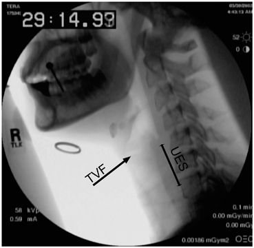
Fig. 5.2
Region of the upper esophageal sphincter (UES) on lateral fluoroscopy. TVF true vocal fold
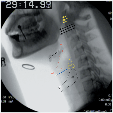
Fig. 5.3
Anatomic structures in lateral fluoroscopic view. Yellow arrows soft palate; Black arrows base of tongue; Black dashed line hyoid bone; TE tip of epiglottis; red dotted line aryepiglottic fold; AC anterior commissure; blue dashed line true vocal folds; LV laryngeal ventricle (space between true and false vocal folds); Yellow dotted line and AC arytenoid cartilage; Black dotted line and CC cricoid cartilage
The PES is made up of the inferior pharyngeal constrictor (IPC), the CPM, and the most proximal cervical esophagus (Fig. 5.1). The actions of all three muscles in addition to the elastic recoil of the thyroid and cricoid cartilages against the cervical spine maintain resting tone. The function of the PES is to prevent aerophagia during respiration and phonation and to protect against aspiration of refluxed gastric and esophageal contents. The PES possesses baseline tone and remains contracted at rest. It reflexively opens during deglutition, eructation (burping), and vomiting. Distension of the esophagus, emotional stress, pharyngeal stimulation, and acid in the esophagus all reflexively tighten the PES. Of the three components that make up the PES, only the CPM contracts and relaxes during all reflex tasks.
Normal PES Opening
The act of swallowing depends upon adequate and timely PES opening . Opening of the PES depends upon muscular relaxation of the CPM , elevation of the larynx, and pharyngeal contraction. Jacob et al. (1989) described five phases of PES opening (Table 5.1) . In the first phase there is an inhibition of tonic PES contraction (Fig. 5.4). This depends on muscular relaxation of the tonically active CPM. Phase I of PES opening is followed by elevation of the hyoid and larynx. Hyolaryngeal excursion (Phase II) provides opening of the PES (Fig. 5.5) through active distraction of the larynx and cricoid cartilages away from the cervical spine. The anterior–superior elevation also helps bring the larynx forward underneath the base of tongue and helps direct the bolus posteriorly toward the hypopharynx . The cartilages do not actually distend off of the spine to open the PES in Phase II, but the region is primed to accept the bolus in preparation for definitive opening in Phase III. The priming provided by elevation appears to be more important than muscular inhibition in Phase I. Elevation is necessary to allow a bolus to enter the PES. Muscular relaxation of the CPM without elevation will not result in PES opening. This has significant clinical implications, as swallowing in individuals with good hyolaryngeal elevation but poor CPM relaxation is possible and often encountered (CPM bar) . Safe and effective swallowing in individuals who can relax their CPM but cannot elevate their larynx off of the cervical spine has not been observed. The advancing bolus will reach a closed PES and follow the path of least resistance into the airway. The third phase of PES opening involves distension of the PES through bolus size and weight (Fig. 5.6). This phase relies upon pharyngeal and lingual peristalsis to propel the bolus past the spacious hypopharynx , through the closed but primed PES, behind the elevating hyolaryngeal complex, and into the cervical esophagus. The elasticity of the elevating PES allows it to be opened by the increasing pressure exerted by the passing bolus. If there is inadequate lingual and pharyngeal contraction, the bolus will not exert enough pressure and the PES will not open. The bolus will again follow the path of least resistance and threaten the airway. The PES cannot adequately be evaluated if lingual and pharyngeal contractility are ineffectual. This is a major limitation of the VFSS , as PES pathology (e.g., CPM bar, stricture, web) cannot be assessed in persons with a significantly weakened tongue or pharynx. In Phase IV of PES opening, the elasticity of the PES causes passive collapse and closure as the bolus passes (Fig. 5.7). The final phase of PES opening, Phase V, involves PES closure through active contraction of the CPM (Fig. 5.8). A disease process that affects any one of the five phases of PES opening can cause the symptom of dysphagia and objective evidence of swallowing dysfunction .
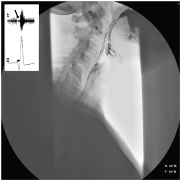
Fig. 5.4
Phase I of UES opening. Inhibition of tonic UES contraction. The baseline electrical activity of the b cricopharyngeus muscle on EMG and B UES pressure on manometry, diminishes (black arrow, asterisk). The barium bolus has been transported to the base of the tongue
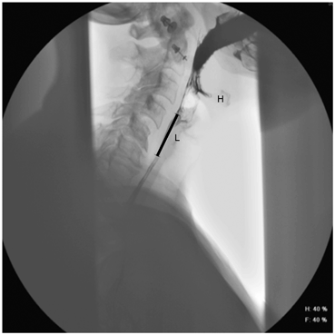
Fig. 5.5
Phase II of UES opening. The larynx (L) and hyoid (H) are elevated, priming the UES for acceptance of the bolus and distension in Phase III. The thyroid cartilage remains apposed to the cervical spine and the UES remains closed (black line)
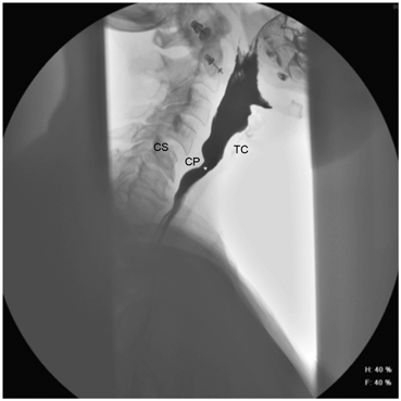
Fig. 5.6
Phase III of UES opening. Distension of the UES by bolus size and weight. This requires adequate lingual and pharyngeal pressure to distract the thyroid cartilage (TC) off of the cervical spine (CS). The UES opens only as little as required to accommodate the advancing bolus (asterisk). CP cricopharyngeus muscle
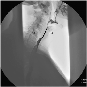
Fig. 5.7
Phase IV of UES opening. The bolus has passed into the esophagus and the elastic recoil of the descending thyroid cartilage (TC) toward the cervical spine (CS) causes passive closure of the UES (black line)
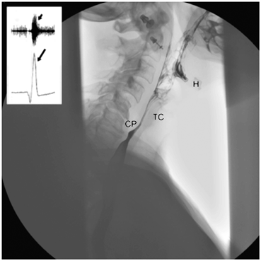
Fig. 5.8
Phase V of UES opening. The hyoid bone (H) and thyroid cartilage (TC) have descended to their pre-swallow positions and the PES closes through active contraction of the cricopharyngeus (CP) muscle. There is post swallow increased electrical activity of the CP muscle on electromyography (short arrow) and a transient elevation of UES manometric pressure above baseline on manometry (long arrow)
Table 5.1
Stages of PES opening
Stage | |
|---|---|
I | Muscular relaxation of the cricopharyngeus muscle |
II | Elevation of the hyoid and larynx |
III | Distension of the PES through lingual and pharyngeal contraction |
IV | Passive closure of the PES through elastic recoil of laryngeal cartilages
Stay updated, free articles. Join our Telegram channel
Full access? Get Clinical Tree
 Get Clinical Tree app for offline access

|