Fig. 4.1
A sagittal rendering of the oral cavity and oropharynx demonstrates normal anatomic structures. CI central incisor; S symphysis of mandible; white arrowhead floor of mouth; white arrow hard palate; OT oral tongue; black arrowhead soft palate; black arrow palatine tonsil; bracket base of tongue (Used with permission from Corey AS, Hudgins PA. Radiographic imaging of human papillomavirus related carcinomas of the oropharynx. Journal of Head and Neck Pathology. 2012 Jul; 6 (1): 25-40.)
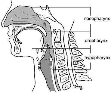
Fig. 4.2
A sagittal drawing demonstrating the three components of the pharynx. (Used with permission from Lee SH. Upper airway structure during development. In: Kheirandish-Gozal L, Gozal D, editors. Sleep disordered breathing in children. New York. Springer Science + Business Media, LLC; 2012)
Some oral and pharyngeal structures including the floor of mouth and lateral pharyngeal walls are not well delineated on fluoroscopy . However a number of osseous and soft tissue components are readily visible and may be reliably assessed with the VFSS (Fig. 4.3). The mandible and maxilla with their associated teeth surround the visible fluoroscopic oral cavity. The oral tongue is best visualized when a radiopaque bolus is in the mouth or is coated with contrast. The soft palate and its most inferior component, the uvula, are generally identifiable as is the tongue base. Posterior to the tongue base, the epiglottis is visible in the air-filled common aerodigestive tract and forms the posterior limit of the vallecula. The posterior portion of the pharyngeal constrictors apposes the cervical spine and easily is assessed.
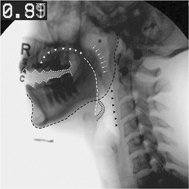
Fig. 4.3
Lateral fluoroscopic view demonstrating important oral and pharyngeal anatomy. White dotted line teeth; black dashed line mandible; white arrowheads oral tongue; white dashed line tongue base; white arrows soft palate; black asterisk nasopharynx; black dotted line epiglottis; black arrowheads posterior pharyngeal wall
Normal anatomic findings include an intact mandibular arch of adequate height with a full complement of healthy teeth. The oral tongue should fill the oral vestibule and the tongue base should be visible beyond the angle of the mandible though not obscuring the vallecula. A perfectly normal epiglottis is thin having a width less than 8 mm or 33 % of the width of C4 (Fig. 4.4). It also follows a slight cranial curvature. The posterior pharyngeal wall should have a near uniform thickness measuring approximately 0.30–0.40 cm between C2 and C3 (Fig. 4.4). Findings outside of these standards may be considered normal and include altered or replaced dentition, mandibular atrophy in the elderly, vallecular filling by the lingual tonsil, retroflexion of the epiglottis, and mild pharyngeal wall thickening particularly if over-lying a prior surgical site or spinal fusion plate. Abnormal fluoroscopic oral and pharyngeal anatomy will be reviewed in Chap. 9.
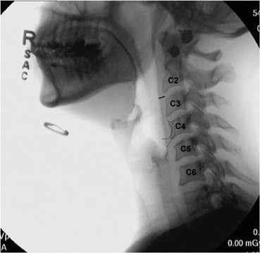
Fig. 4.4
Lateral fluoroscopic view showing normal posterior pharyngeal wall (black line) and epiglottic thickness. An outline of the epiglottis (grey dotted line) has been superimposed to demonstrate that its width is less than 33 % of C4
Normal Fluoroscopic Physiology
VFSS affords the unique ability to evaluate the posterior oral portion of deglutition, which remains hidden during clinical swallow evaluations and flexible endoscopic examination of swallowing (FEES). The VFSS is helpful in correlating clinical oral motor findings with bolus control.
Stay updated, free articles. Join our Telegram channel

Full access? Get Clinical Tree


