Normal Anatomy and Histology, Specimen Processing, Pathologic Reporting, and Artifacts
Allen P. Burke, M.D.
Joseph J. Maleszewski, M.D.
Pulmonary Anatomy and Histology
General Features and Development
The respiratory system broadly includes the acini, which are primarily responsible for gas exchange, and the airways and blood vessels that deliver the gases and blood, respectively, to such.
The lungs begin development as bilateral and symmetric structures; they acquire asymmetry through development and therefore ultimately exhibit sidedness (situs). Pulmonary sidedness is determined by the position of the morphologic right and left lungs, which is largely driven by the relative position of the pulmonary arteries and bronchi. In normal sidedness (situs solitus), the right mainstem bronchus is short and eparterial, meaning that the right pulmonary artery travels anterior to the right upper and intermediate bronchi. The left upper lobe bronchus is longer and hyparterial, passing inferior to the left pulmonary artery (Fig. 1.1).
Airways
Airways can be categorized by structure/size (large cartilaginous and small noncartilaginous) and by function. The latter categorization divides airways into those that are responsible for transmitting gas to and from the units of gas exchange (conducting airways) and those that actually contain gas exchange units (respiratory airways). Regardless of classification, the airways begin at the trachea and end at the respiratory bronchiole.
The trachea enters the thoracic inlet, just distal to the larynx, along with vascular structures, esophagus, muscles, vagus and phrenic nerves, and thoracic duct. The trachea divides into the right and left bronchi, with a more acute angle to midline on the right (20 degrees) than on the left (35 degrees), leading to a propensity for aspirated material to enter the right bronchus. In addition to transmitting air to and from the acini, they also provide an important protective role, both immunologic and physical, with their lymphoid, epithelial, and mucociliary structures.
The airways are lined by respiratory epithelium, under which is the muscular layer. The bronchi have a cartilaginous and fibrous layer under the muscularis and mucus glands between the muscularis and cartilage (submucosa). The respiratory epithelium consists of mucussecreting goblet cells, ciliated cells, scattered neuroendocrine cells, and a basal layer. Additionally, there is a population of pulmonary brush cells, which differ ultrastructurally from ciliated cells, thought to be involved in fluid absorption.1 The ciliated cells are important in mucous transit and the clearance of particulate matter from the airways back to the environment. Ultrastructurally, the cilia consist of nine doublets that surround a central pair (Fig. 1.2). Dynein arms (inner and outer) join the peripheral doublets, and radial spokes connect the peripheral doublets to the central pair. Identification of these normal structures is critical in the evaluation for primary ciliary dyskinesia.
All epithelial cells express cytokeratins, including cytokeratin 7; in addition, the basal cells express p40 and p63. TTF-1 expression is limited to respiratory bronchioles, which contain Clara cells (surfactantproducing cells), and to alveolar lining cells (pneumocytes). In general, the number of goblet and ciliated cells decreases in the distal airways, and the respiratory bronchioles are composed primarily of basal and Clara cells.
Lobation and Lung Segments
The left and right lung lobes are separated by interlobar fissures, usually one on the left and two on the right (Fig. 1.1). Oblique fissures, on each right and left lung, divide the upper and lower lobes and travel from the upper lateral to lower medial lungs. A horizontal fissure on the right separates the upper and middle lobes. Incomplete development or absence of the horizontal fissure is a common variant, resulting in a two-lobed right lung.2 This is why lung laterality is best assigned by the relationship of the bronchus and pulmonary artery, rather than lobation.
There are 19 bronchopulmonary segments: 10 on the right and 9 on the left, owing to fusion of the apical and posterior segments of the left upper lobe (Table 1.1). Secondary pulmonary lobules (Fig. 1.3), the smallest unit recognized by high-resolution computed tomography (CT), are 1.0- to 2.5-cm polyhedral collections of acini (see below) served by terminal bronchioles. They are bounded by the pleura and the interlobular septa.
Alveoli
Each secondary lobule contains between 10 and 15 acini (the functional units), which include all the alveoli containing structures distal to the terminal bronchiole. The alveoli are the sac-like structures involved in gas exchange and receive air from the upstream airways. The alveoli themselves measure ˜200 µm across. Some alveolar sacs arise directly from respiratory bronchioles without connections through primary lobules.
The histologic features in two dimensions do not readily allow for distinction between the various alveolar compartments. Respiratory bronchioles may be seen adjacent to alveoli, whereas bronchioles are generally seen on cross section.
The lining cells of the terminal bronchioles and alveoli are composed of TTF-1-positive Clara cells in the former and pneumocytes in the latter. Mature type I pneumocytes are flattened, attenuated squamous cells with abundant cytoplasm, and small nuclei are generally not visible in normal sections and cover over 95% of the alveolar surface. They are attached to one another by desmosomes and occluding junctions. Pneumocytes with the ability to regenerate are type II pneumocytes, which have surfactant production capability. They are cuboidal and cover a much smaller surface area (<5%), despite the fact that they are actually more numerous than type I pneumocytes.
Approach to Histologic Evaluation of Peripheral Lung Tissue
When evaluating lung tissue, starting at low magnification helps to characterize the overall architecture and pattern of any pathology. Identification of bronchovascular bundles, small airways, interlobular septa, alveolar septa, and pleural surfaces should also be done at this
time (Figs. 1.4, 1.5, 1.6, 1.7). Patterns of pathology can help to establish a differential diagnosis.
time (Figs. 1.4, 1.5, 1.6, 1.7). Patterns of pathology can help to establish a differential diagnosis.
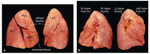 FIGURE 1.1 ▲ Gross pulmonary anatomy. A. Normal lobation pattern. B. Normal relationship of the pulmonary artery and bronchus. A morphologic right lung shows the pulmonary artery anterior to the upper lobe bronchus, while a morphologic left lung shows the pulmonary artery superior to the upper lobe bronchus. |
Bronchiolocentric processes occur around the bronchioles, while angiocentric lesions occur around the adjacent muscular pulmonary arteries. Septal and paraseptal patterns of disease/injury follow the interlobular septa that bound the pulmonary lobules. Lymphatics are present along the pleura, bronchovascular bundles, and interlobular septa, which will all tend to be involved in diseases with a lymphatic distribution. Consolidative processes will fill the airspaces and make the lung look more “solid.” Finally, diffuse interstitial diseases can involve virtually all of the nonairspace regions of the pulmonary microanatomy. Although the limits of the secondary lobules cannot usually be clearly seen in normal lung, the parenchyma around the bronchovascular bundles is generally centrilobular or centriacinar and that alveoli near pleural surfaces or lobular septa are peripheral.
 FIGURE 1.2 ▲ Ciliary ultrastructure. Nine outer doublets radiate around a central pair and are connected to such via radial spokes. Inner dynein arms (IDA) and outer dynein arms (ODA) are visible extending from the outer doublets. Absences or abnormalities of these dynein arms are the most common morphologic finding in cases of primary ciliary dyskinesia. |
Pulmonary Arteries
The pulmonary trunk branches into the right and left pulmonary arteries. The right pulmonary artery, longer than the left, travels beneath the aortic arch before entering the lung hilum. The arteries divide into lobar and segmental branches, with names similar to the bronchopulmonary segments that they feed. The bronchial arteries arise from the thoracic aorta just distal to the arch, directly either from the aorta or from the intercostal arteries, and usually with one on the right and two on the left.
Histologically, pulmonary arteries are identified adjacent to the bronchi and in bronchovascular bundles toward the periphery. Normally, there is little intima, and the media is thin (10% or less of the diameter of the artery) (Fig. 1.8). There may be concentric intimal thickening (or hyalinosis) as a result of aging or increased arterial pressures. Typically, the artery is similar in diameter to the accompanying airway. The proximal arteries accompanying the bronchi (main pulmonary arteries and lobar arteries) are elastic, with concentric elastic lamellae, seen best on elastic stains. There is a gradual transition to muscular arteries accompanying segmental bronchi and bronchioles.
Muscular pulmonary arteries have a distinct internal and external elastic lamina, unlike bronchial arteries, which tend to have only a distinct internal elastic membrane with a more fragmented external elastic membrane (like all other systemic muscular arteries).
Muscular pulmonary arteries have a distinct internal and external elastic lamina, unlike bronchial arteries, which tend to have only a distinct internal elastic membrane with a more fragmented external elastic membrane (like all other systemic muscular arteries).
TABLE 1.1 Lung Segments | ||||||||||||
|---|---|---|---|---|---|---|---|---|---|---|---|---|
|
Stay updated, free articles. Join our Telegram channel

Full access? Get Clinical Tree


