Nonoperative Management of Chronic Venous Insufficiency


Nonoperative Management of Chronic Venous Insufficiency


James R. Ballard and Gregory L. Moneta


Chronic venous insufficiency (CVI) is common, ranging from primary varicose veins to valvular incompetence of the lower extremity veins. Most symptoms of CVI can be improved with bedrest and elevation of the legs. However, prolonged immobilization is impractical and dangerous for most patients. Nonoperative therapy remains the standard initial treatment for CVI. The goal of nonoperative therapy in CVI is to control symptoms and maintain a patient’s functional ability without the need for prolonged bedrest or for surgery. Nonoperative treatment strategies for CVI despite progress in ablative and reconstructive venous surgery.


Nonoperative treatment strategies for CVI consist of mechanical and pharmacologic therapies. External compression therapy is generally regarded as primary therapy for all forms of CVI, including venous ulcer. Pharmacologic therapy serves primarily as an adjunct to compression therapy in patients with relatively mild manifestations of CVI. It is seldom used as sole therapy for treatment of CVI and is employed more often in Europe than in the United States.


Compression Therapy


Ambulatory venous hypertension acts through largely unknown mechanisms to facilitate the tissue damage characteristic of CVI. The effects of increased ambulatory venous hypertension must be overcome for CVI therapy to be effective. The pressure in a leg vein reflects the weight of the blood column between the site of measurement and the right atrium. In the supine position, venous pressure is between 10 and 20 mm Hg. With a patient in the supine position, the lower leg veins are narrowed by an external pressure of 10 to 20 mm Hg and occluded by a pressure of 20 to 25 mm Hg. In the upright patient, depending on the patient’s height, the pressure in the lower leg is approximately 60 mm Hg. Upright, an external pressure of 35 to 40 mm Hg can narrow lower extremity veins. Pressures greater than 60 mm Hg are required to occlude the lower leg veins while in the standing position. Given this, major hemodynamic effects of external compression require a pressure of 35 to 40 mm Hg. Microcirculatory studies show the safe upper limit is 60 mm Hg for sustained compression however, intermittent pressure peaks can exceed this upper limit safely.


External compression improves venous pump function in patients with CVI (CEAP 3–6) and depends on the interface pressure. Inelastic bandages do not give way to changes in leg circumference. They produce a more-pronounced reduction of venous reflux compared to elastic materials applied with the same initial resting pressure. In patients with CVI who walked on a treadmill, inelastic bandages applied with a resting pressure greater than 50 mm Hg significantly decreased ambulatory venous pressure measurements. Inelastic bandages produce intermittent occlusion of the leg veins by peak pressures of 80 mm Hg during walking. Elastic stockings only increase interface pressure by 3 to 8 mm Hg higher than the resting pressure.


The exact local physiological and biomechanical mechanisms by which compression therapy improves symptoms of CVI and healing of venous ulcerations remain uncertain. The target organ of CVI is the skin. Compression therapy provides multiple potential benefits to the skin and subcutaneous tissue. Microcirculatory hemodynamics may be favorably altered by compression therapy. Supine perimalleolar subcutaneous pressures increase with elastic compression. This can produce a Starling gradient favoring movement of fluid from the interstitial space to the lymphatic circulation, counteracting the forces favoring leakage of fluid from the capillary. These observations are concordant with the fact that elastic and nonelastic bandages reduce lower extremity edema in patients with CVI. With edema reduction, cutaneous and subcutaneous metabolism can improve from enhanced diffusion of oxygen and other nutrients to the skin and subcutaneous tissues.


A number of biomechanical abnormalities have been implicated in venous ulceration. Tumor necrosis factor α (TNF-α) and vascular endothelial growth factor (VEGF) likely participate in the tissue damage seen in patients with CVI. Venous stasis ulcers treated with compression therapy for 4 weeks have been shown to have decreased levels of these cytokines, and the decreases correlated with the degree of ulcer healing.


Several etiologies exist for chronic leg swelling as well as for leg ulcers. In the majority of cases, chronic leg swelling is not a result of venous disease, and only 70% of leg ulcers are venous in origin. A definitive diagnosis of CVI and/or venous ulceration is necessary before initiation of treatment with compression therapy. A number of medical conditions and medications can promote lower extremity edema and/or ulceration. Severe arterial insufficiency must be excluded by noninvasive studies before therapy for CVI is initiated. Venous ulceration in the setting of coexisting arterial insufficiency, especially severe ulceration, is difficult to heal with compression and is theoretically dangerous.


Elastic Stockings


Gradient elastic compression stockings are the most common method for delivering compression therapy. Initially developed in the 1950s, they were made to simulate gradient hydrostatic forces similar to the forces exerted by water in a swimming pool. Gradient elastic compression stockings are manufactured worldwide and are readily available in various strengths, lengths, and compositions.


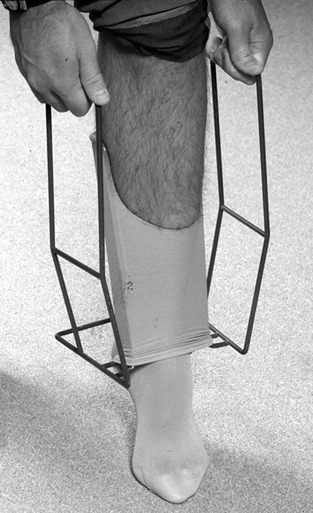
One advantage of elastic stockings for treating CVI and/or venous ulceration is that their effects are operator independent. As long as the patient wears the stockings, the effects are dependent on the strength of the stocking and independent of the patient. Compression stockings are less bulky than compression bandages and potentially more comfortable. They may be worn with normal footwear and allow daily inspection of ulcers as well as wound care. Because they can be difficult to apply, adjunctive devices are often useful (Figure 1).


Paste Boots


Paste boots are a form of compression developed by German dermatologist Paul Gerson Unna in 1896. Unna’s boot has been used for decades to treat venous ulcers. There are multiple versions of the basic concept of the Unna boot. The typical Unna’s boot dressing consists of three or four layers. It is best applied by trained personnel. The first layer is rolled gauze bandage impregnated with calamine, zinc oxide, glycerin, sorbitol, gelatin, and magnesium silicate. It is applied with graded compression from the forefoot to just below the knee. Additional layers consist of continuous gauze dressings. The outer layer is a wrap with graded compression also.


The bandage stiffens with drying, resulting in rigidity that can help prevent and help resolve edema. Pressure in the distal lower leg in the supine position following application may be 50 to 60 mm Hg. The dressing requires weekly changing unless there is significant drainage. Advantages of Unna’s boot include minimal patient involvement, continuous compression, and significant effectiveness in healing ulcers. In a 15-year review of 998 patients with venous ulcers treated with Unna’s dressings, 73% of ulcers healed in patients who returned for more than one treatment. Median time for healing for individual ulcers was 9 weeks.


Unna’s dressings have several disadvantages. Their placement can be time consuming for the provider to apply, and the ulcer cannot be monitored daily. The dressing itself is bulky and uncomfortable for some patients. The amount of compression is operator dependent, and patients can develop contact dermatitis.

Stay updated, free articles. Join our Telegram channel

Aug 25, 2016 | Posted by in CARDIOLOGY | Comments Off on Nonoperative Management of Chronic Venous Insufficiency

Full access? Get Clinical Tree

Get Clinical Tree app for offline access