Noninfectious Conditions in Patients with Human Immunodeficiency Virus Infection

Chapter 30 Noninfectious Conditions in Patients with Human Immunodeficiency Virus Infection


Although most complications affecting the lungs during the course of HIV infection arise from the viral infection itself, especially as reported early in the epidemic, HIV-infected patients may present with or subsequently develop complications related to alterations in immune regulation. A marked CD8+ T cell infiltrate in the lung may be caused by HIV in both symptomatic and nonsymptomatic patients. The effects of the virus on the pulmonary microenvironment include a progressive decline in local immunocompetence that results in failure to mount a protective immune response against opportunistic infections. The spectrum of noninfectious complications associated with HIV infection encompasses other idiopathic conditions and pulmonary malignancies, which include Kaposi sarcoma, Hodgkin and non-Hodgkin lymphomas, and solid tumors (Table 30-1 and 30-2).


Table 30-1 Noninfectious Pulmonary Conditions Associated With Human Immunodeficiency Virus Type 1 Infection


Disease Category Specific Disease(s)
Neoplastic disease
Inflammatory disease
Airway disease
Pulmonary vascular disease Pulmonary hypertension
Miscellaneous Antiretroviral treatment–induced respiratory disease

Table 30-2 Association of Noninfectious Pulmonary Conditions with Degree of Immunodeficiency


Degree of Immunodeficiency Noninfectious Pulmonary Condition
Severe (CD4+ cell count less than 50 cells/µL)
Moderate to severe (CD4+ cell count 50-200 cells/µL)
Moderate (CD4+ cell count 200-350 cells/µL)
Normal (CD4+ cell count greater than 350 cells/µL)


Neoplasia


Cancer incidence is higher in persons with immunodeficiency of all causes. In HIV-1 infection, cancers that are uncommon in the general population predominate and include mainly lymphoma and Kaposi sarcoma. Other, more common cancers are now seen with increasing frequency; however, as HIV-1 infection has become controllable with antiretrovirals.


Kaposi Sarcoma


Epidemiology, Risk Factors, and Pathogenesis


In the general population, Kaposi sarcoma is a rare benign skin tumor that develops in the skin of the lower extremities in elderly men from the Mediterranean area. The disease, originally described in 1872 by the Hungarian physician Moricz Kaposi, is 100 to 1000 times more frequent in persons with HIV-1 infection than in the general population and is associated with an aggressive course. The incidence of AIDS-associated Kaposi sarcoma has, however, declined substantially over the past 2 decades. Before the advent of cART, incidence rates ranged from 25 to 50 cases per 1000 person-years of follow-up (PYFU) but have steadily declined, so that the incidence in a cART-treated population is less than 5 per 1000 PYFU. Approximately 5% to 10% of all diagnosed cases of AIDS are based on a finding of Kaposi sarcoma. HIV-1–infected men who have sex with men have a 20 times higher risk for development of this tumor than in persons in other transmission groups. Frequency increases with lower CD4+ T cell counts, but the disease can occur at all levels of immunodeficiency. Thus, when Kaposi sarcoma is the AIDS-defining illness, it may occur at a relatively higher CD4+ T cell count than other such illnesses.


Kaposi sarcoma is an angioproliferative inflammatory condition that is associated with infection with human herpesvirus type 8 (HHV-8). Gene expression profiling shows that the lesions consist of aberrant endothelial and inflammatory cells of lymphatic origin. HHV-8, a γ-2 herpesvirus of the Rhadinovirus genus, has been demonstrated in all forms of Kaposi sarcoma, and in situ hybridization has demonstrated the presence of HHV-8 RNA and DNA in spindle, endothelial, and mononuclear cells of the tumor. HHV-8 DNA is detectable in peripheral blood before the development of the tumor, and increased viral replication is evident before onset of symptoms. More than 80% of patients with AIDS-associated Kaposi sarcoma have antibodies to HHV-8. HHV-8 may exert its oncogenic potential in several ways. The HHV-8 genome contains several genes that are homologues to human genes. Among these is a latent nuclear antigen that binds to p53 and associates viral DNA to human DNA during mitosis; a viral cyclin that activates cyclin-dependent kinases to prevent human cells from remaining in a G1 phase; a constitutively expressed receptor (viral interleukin-8 receptor) that may be involved in angiogenesis; a Bcl-2 like protein that prevents apoptosis; and viral homologues to cytokines (viral macrophage inflammatory protein [MIP] and viral interleukin-6) that may be responsible for some of the constitutive symptoms associated with Kaposi sarcoma. Cytokines are required for HHV-8–infected endothelial cells to acquire their phenotype and for continued growth of the tumor. The HIV-1 Tat protein upregulates cytokines and metalloproteinases that further promotes the oncogenic potential of Kaposi sarcoma cells.


Clinical Features


Cutaneous and mucocutaneous manifestations of AIDS-associated Kaposi sarcoma are the more common and usually precede visceral disease by months to years. Skin and visceral lesions appear as red or violet macules, papules, or nodules that may coalesce to form plaque-like lesions. Lesions may affect any area of the skin and involve any organ system. Lymphadenopathy is frequent.


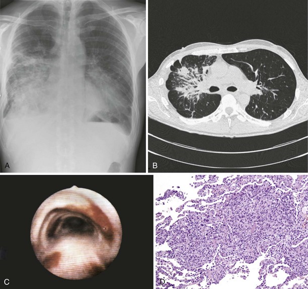
Pulmonary Kaposi sarcoma may cause nonproductive cough, hemoptysis, shortness of breath, chest pain, and fever. In rare instances, involvement of the larynx or trachea may cause airway obstruction. Extrapulmonary involvement is frequent, but 15% of cases of Kaposi sarcoma occur without skin lesions. CD4+ cell counts are low (0 to 100/µL) at the time of diagnosis. Examination of the lungs usually shows no abnormalities. Chest radiograph may be normal in appearance but commonly shows singular or multiple peribronchovascular nodules (Figure 30-1, A and B). Diffuse infiltration and air space consolidation also may be present. Pleural effusion is common. Hilar and mediastinal lymphadenopathy may be visible on chest films but are better visualized with computed tomography (CT) scanning. With pulmonary disease, CT scans typically reveal peribronchovascular nodules that are larger than 1 cm in diameter. The role of magnetic resonance imaging (MRI) or positron emission tomography (PET) in the diagnosis and management of pulmonary Kaposi sarcoma is not clear.


Diagnosis


Pulmonary Kaposi sarcoma is highly likely in a HIV-1–infected person with a CD4+ cell count of less than 100/µL, the characteristic cutaneous or mucocutaneous lesions, and pulmonary symptoms. Bronchoscopy is useful to visualize typical endobronchial lesions (see Figure 30-1, C). These are flat or slightly raised and occur throughout the tracheobronchial tree but are seen most frequently at airway bifurcations. Pleural effusions usually are exudative and may be serous or serosanguineous. Serum lactate dehydrogenase may be elevated. Histologic verification is usually not required for a diagnosis of Kaposi sarcoma. Endobronchial biopsy may confer a substantial risk of severe bleeding, but biopsy may be necessary in cases in which bronchial involvement is lacking. In such cases, transbronchial or percutaneous needle biopsy is useful. In cases of pleural Kaposi sarcoma, video-assisted thoracoscopy can be used to visualize pleural lesions and to perform biopsy. Open lung biopsy in this setting is obsolete because of the associated complications. Lung biopsy specimens show typical features of Kaposi sarcoma, which include a tumor-like infiltrate with a peribronchovascular distribution of spindle cells. Slitlike spaces without endothelium contain extravasated erythrocytes (see Figure 30-1, D). In situ hybridization and immunostaining usually are positive for HHV-8. HHV-8 DNA is detectable in bronchoalveolar lavage fluid (BAL) cells.


Treatment


All patients with Kaposi sarcoma should be treated with cART, but immune reconstitution may induce an inflammatory reaction and flare of the disease within 2 to 8 weeks of initiation of this therapy. Cutaneous and mucocutaneous lesions of AIDS-associated Kaposi sarcoma usually regress with cART without chemotherapy but may require additional local radiotherapy. Visceral Kaposi sarcoma, however, will necessitate chemotherapy. Concomitant use of cART increases the response rate to chemotherapy. All patients should be offered P. jirovecii prophylaxis regardless of their CD4+ cell count. Pegylated liposomal doxorubicin or liposomal daunorubicin are used for first-line treatment of Kaposi sarcoma, in addition to cART. In randomized multicenter trials, response rates and adverse effect profiles to both agents given as monotherapy proved to be superior to conventional chemotherapy.


Liposomal anthracyclines are well tolerated overall. Myelosuppression is the most important dose-limiting toxicity. The risk of drug-drug interactions with antiretroviral agents is low for anthracyclines. The taxane paclitaxel, in combination with granulocyte colony-stimulating factor (G-CSF), is approved for second-line treatment of Kaposi sarcoma. Experience with paclitaxel and pulmonary Kaposi sarcoma is limited, and the use of paclitaxel is associated with more severe adverse effects than those reported with anthracyclines. Paclitaxel has potential serious drug-drug interactions with antiretroviral drugs.


Due to the high cost of anthracyclines and taxanes their use may be limited in resource-poor settings. Alternatively, the most widely used regimen for the treatment of pulmonary Kaposi sarcoma, a combination regimen of doxorubicin (Adriamycin), bleomycin, and vincristine (ABV) may be used. Vincristine may be replaced with vinblastine in patients with polyneuropathy. The combination is given every 2 weeks. Response is evaluated after four to six series of chemotherapy. Response rates are 30% to 50%. Adverse side effects include anemia, neutropenia, thrombopenia, neuropathy, mucositis and alopecia. There are potential drug-drug interactions between vincristine or vinblastine and antiretroviral agents.


Investigational agents include thalidomide, imatinib, mTOR inhibitors, IL-12, and fumagilin.


Jun 12, 2016 | Posted by in RESPIRATORY | Comments Off on Noninfectious Conditions in Patients with Human Immunodeficiency Virus Infection

Full access? Get Clinical Tree

Get Clinical Tree app for offline access