Nonbacterial Infectious Pneumonia

Chapter 26 Nonbacterial Infectious Pneumonia


Nonbacterial causes of infectious pneumonia are important diagnostic considerations in the evaluation of patients who are immunocompromised, who have recognized structural lung disease, or who do not respond to appropriate antibiotic therapy for presumed bacterial pneumonia. A detailed clinical history can assist the astute clinician in making the diagnosis of a nonbacterial cause of infectious pneumonia in these circumstances. Important information to gather includes travel history, geographic location, occupational history, and recreational activities. This chapter presents an overview of common fungal and parasitic causes of pneumonia, including clinical presentation, diagnostic considerations, and treatment options.


Fungal Pneumonias


Aspergillosis


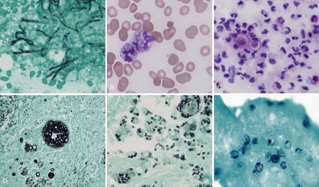
Aspergillus organisms are ubiquitous saprophytic fungi. They grow well in soil and decaying vegetation. These fungi also have been found in hospitals, ventilation and water systems, and dust associated with construction activity. Disease manifestations depend on the immune status and lung structure of the host. Clinical manifestations include allergic disease, airway colonization, aspergilloma formation, tracheobronchial disease, chronic necrotizing pneumonia, and invasive disseminating disease. Allergic disease generally is seen in immunocompetent patients and may encompass airway hyperreactivity, allergic bronchopulmonary aspergillosis (ABPA), and hypersensitivity pneumonitis. Airway colonization occurs in patients with impaired mucociliary clearance or distorted lung structure such as in bronchiectasis. Aspergillomas, or “fungus balls,” thrive in cavitary lung lesions such as those associated with tuberculosis. Tracheobronchial aspergillosis and chronic necrotizing pneumonia (locally invasive aspergillosis) are manifestations found in patients with acquired immunodeficiency syndrome (AIDS), recipients of transplanted organs (especially lungs), and patients with obstructive lung disease who use inhaled or systemic steroids. Finally, invasive or disseminated aspergillosis develops in the context of profound and protracted granulocytopenia (Figure 26-1).


Fungal culture and histopathologic examination constitute the “gold standard” for the diagnosis of aspergillosis. The diagnosis has been simplified by the advent of the galactomannan detection assay, which confers a high degree of sensitivity and specificity. The treatment of aspergillosis is tailored to the disease manifestation. Allergic manifestations are managed with routine asthma care that includes avoidance of allergen exposures and maintenance bronchodilators. Steroids are reserved for severe cases. ABPA mandates treatment with corticosteroids initially and may require the addition of itraconazole. Aspergillomas can be followed clinically and generally remain dormant but can manifest with massive hemoptysis necessitating embolization or surgical resection. Voriconazole is the preferred agent for treatment of invasive aspergillosis. Liposomal amphotericin B, posaconazole, and echinocandins are reserved for salvage therapy.


Histoplasmosis


Histoplasmosis is the most prevalent endemic mycosis in North America. Histoplasma capsulatum is a dimorphic fungus existing as a mold in nature. It forms spores that become aerosolized, facilitating inhalation. After inhalation, H. capsulatum grows as yeast forms in the host.


The clinical manifestations of histoplasmosis are variable and depend on the intensity of exposure along with the immune status and underlying lung architecture of the host. The Mississippi and Ohio River valleys are highly endemic. Histoplasmosis also is endemic to parts of Central America, South America, Africa, and Asia. Moist soil is an ideal habitat for Histoplasma, especially when supplemented with bird or bat guano. Activities that can lead to infection include spelunking (caving), excavation, demolition, and cleaning of chicken coops or old buildings. After inhalation, the spores are converted into yeasts, which are phagocytosed by macrophages. The organism is able to survive and proliferate inside macrophages, allowing it to disseminate. Within 2 weeks, however, a protective cellular immune response usually develops, which contains the fungus by forming granulomas (often seen as calcified granulomas on chest radiographs). Most otherwise healthy persons infected with a low inoculum remain asymptomatic (i.e., the infection is subclinical) or are minimally symptomatic. Patients with impaired immunity that fails to contain the infection can develop progressive disseminated histoplasmosis, with involvement of the bone marrow, liver, spleen, adrenal glands, gastrointestinal tract, and central nervous system (CNS). This manifestation typically is seen in persons with AIDS or patients taking immunosuppressive medications such as prednisone, methotrexate, and anti-TNF-α agents. Common clinical manifestations of disseminated disease include fever, weight loss, hepatosplenomegaly, pancytopenia, meningitis, focal brain lesions, ulcerations of the oral mucosa, gastrointestinal ulcerations, and adrenal insufficiency. Disseminated histoplasmosis carries a high mortality rate unless promptly diagnosed and appropriately treated (Table 26-1).
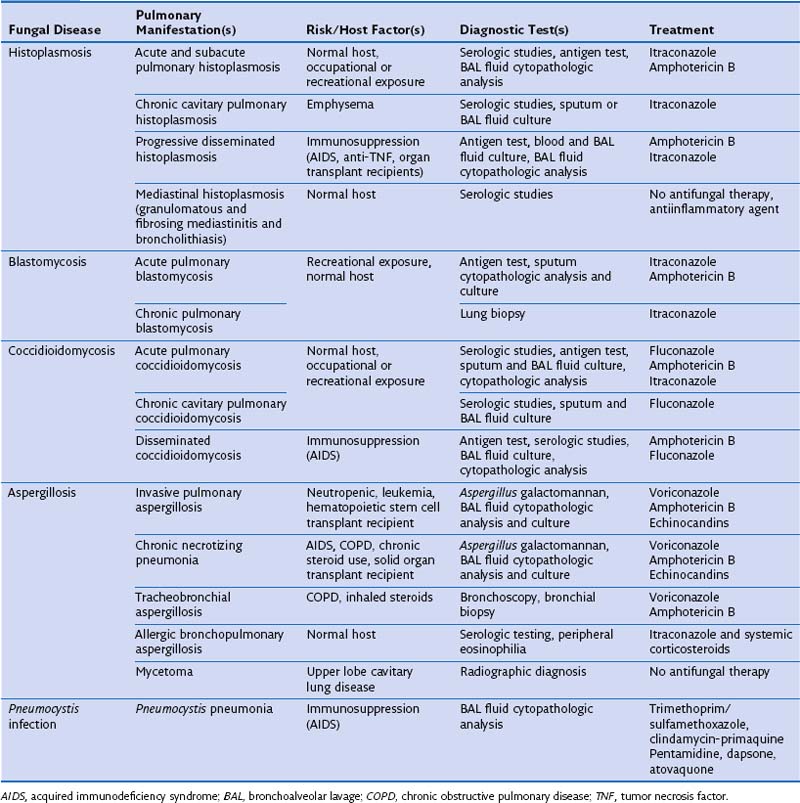

Patients who inhale larger inocula of Histoplasma conidia during outbreaks or in an enclosed space may develop severe pulmonary disease with a flulike illness that can lead to respiratory failure. When the infection occurs in the context of structural lung disease (bullous emphysema), it may not be effectively contained, and patients can develop chronic cavitary histoplasmosis with upper lobe infiltrates, cavitations, and pleural thickening. Other late manifestations of histoplasmosis include fibrosing mediastinitis (a late complication associated with high morbidity), calcification of mediastinal nodes, and broncholithiasis.


Identification of H. capsulatum is accomplished using a combination of tests. Fungal culture remains the “gold standard.” However, H. capsulatum may require up to 4 weeks to be grown in culture, limiting its utility in severe cases. Rapid diagnostic testing using antigen detection in serum or urine samples is useful in patients with moderate to severe disease. Antigen detection in bronchoalveolar lavage (BAL) fluid may help make the diagnosis of pulmonary histoplasmosis. Rapid diagnosis also can be achieved through cytopathologic examination, albeit with lower sensitivity than that attained with culture or antigen detection. Histoplasma serologic testing (immunodiffusion and complement fixation) is particularly useful in chronic (cavitary) and subacute disease, because detectable levels of antibody appear after 4 weeks of infection.


Treatment for histoplasmosis is reserved for patients with chronic pulmonary, disseminated, or acute moderate to severe disease. Liposomal amphotericin B followed by itraconazole is the preferred regimen for severe cases. Mild to moderate disease can be treated with itraconazole exclusively. Corticosteroids also may be useful in cases of severe, life-threatening acute pulmonary histoplasmosis. The duration of treatment for acute pulmonary histoplasmosis is 6 to 12 weeks, whereas that for disseminated and chronic pulmonary histoplasmosis is 12 to 18 months.


Fibrosing mediastinitis and broncholithiasis represent chronic reactions to past infection and do not respond to antifungal therapy. Treatment consists of endoscopic or surgical removal of partially or completely eroded broncholiths. Placement of endovascular stents may be required in advanced fibrosing mediastinitis causing obstruction of major mediastinal vessels. Obstruction of central airways or the esophagus also can occur and may necessitate balloon dilatation or stenting.


Blastomycosis


Blastomyces dermatitidis is a dimorphic fungus that causes pulmonary and systemic disease. Most cases of blastomycosis have been reported in North America, where it occurs predominantly in the Midwestern states and Canadian provinces surrounding the Great Lakes. In the United States, the endemic region overlaps significantly with that of H. capsulatum along the Mississippi and Ohio River valleys. Cases have been reported from Africa and India as well. When outbreaks occur, they usually are associated with outdoor recreational activities or work around waterways. As with histoplasmosis, the mechanism of infection involves inhalation of the spores, which are then converted to the yeast form in the lung; however, polymorphonuclear cells are abundant in blastomycosis lesions, mimicking pyogenic infections.


Blastomycosis can manifest clinically in a variety of ways. The most common presentation is pulmonary disease, followed by involvement of the skin, bone, joints, and CNS. Acute pulmonary blastomycosis resembles community-acquired pneumonia and often is mistakenly treated for bacterial pneumonia before the correct diagnosis is made. Signs and symptoms include fever, chills, productive cough, and chest pain. Radiographic findings include consolidation, cavities, nodules, and miliary patterns. Mediastinal adenopathy and pleural effusions are uncommon. Acute blastomycosis can progress to acute respiratory distress syndrome (ARDS), even in immunocompetent patients. Risk factors for severe infection include diabetes and diffuse pulmonary involvement. Chronic pulmonary blastomycosis mimics lung cancer or tuberculosis and can be insidious in onset and progression and minimally symptomatic. Skin lesions often are mistaken for squamous cell carcinoma or pyoderma gangrenosum.


Definitive diagnosis requires growth of Blastomyces from sputum, BAL fluid, or biopsy material. Rapid identification is made by visualization of the characteristic broad-based budding yeast in clinical specimens. Typically, blastomycosis is diagnosed when a presumed community-acquired bacterial pneumonia fails to respond to antimicrobial therapy or if a mass suspicious for malignancy is detected. When the clinical presentation includes cutaneous manifestations, the diagnosis can be secured by skin biopsy. Blastomyces antigen can be found in the urine, serum, or BAL fluid, providing a reliable means for establishing a rapid diagnosis, especially in severe cases. The current antigen assays do not differentiate histoplasmosis from blastomycosis, as a consequence of cross-reactivity. Histopathologic and cytopathologic analysis can be helpful in differentiating histoplasmosis from blastomycosis and should be routinely ordered with invasive testing procedures.

Stay updated, free articles. Join our Telegram channel

Jun 12, 2016 | Posted by in RESPIRATORY | Comments Off on Nonbacterial Infectious Pneumonia

Full access? Get Clinical Tree

Get Clinical Tree app for offline access