Non-Small Cell Lung Cancer
Presentation
An 87-year-old woman is referred to you with complaints of blood-streaked sputum for the past 3 weeks. The patient reports a productive cough for several months. She also reports a two-pack-per-day smoking history for the past 15 years.
Past surgical history is pertinent for a previous hiatal hernia repair. On review of systems, the patient denies any current heartburn or dysphagia but does admit to a 5-pound weight loss and recent upper back pain.
On physical examination, the breath sounds are diminished at the bases, and there is wheezing present over the right chest. In addition, there is focal chest wall tenderness over the right lower ribs posteriorly.
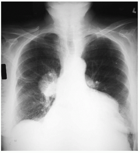
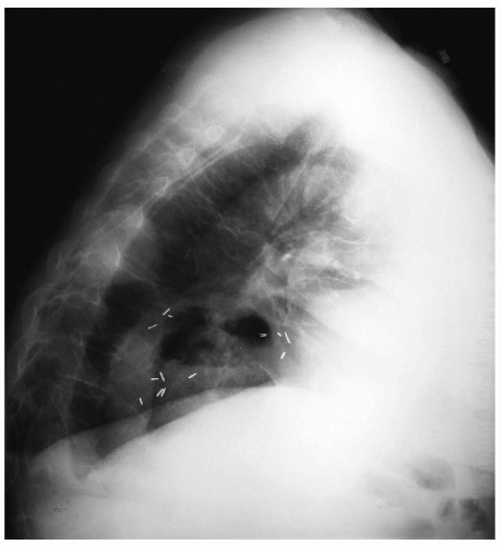
The following x-rays are obtained.
Differential Diagnosis
Hemoptysis usually indicates the presence of infectious or neoplastic lesions of the lung. Given this patient’s strong smoking history and the presence of an irregular density on chest x-ray, a lung malignancy is the most likely diagnosis.
Recommendation
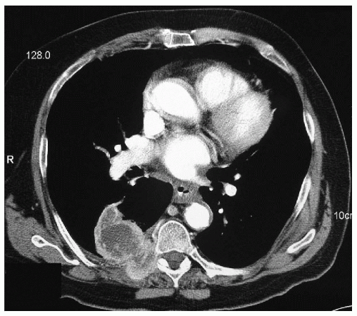
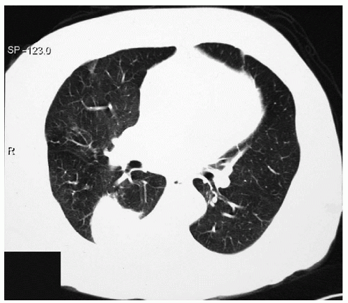
Computed tomography (CT) scans of the chest are needed to define the location and extent of the mass and to aid in staging.
▪ CT Scans
CT Scan Report
There is a large, necrotic, pleural-based mass in the right lower lung posteriorly. This mass, which measures 8 cm × 5 cm, invades the adjacent rib and extends into the paraspinous musculature. Nonspecific parenchymal opacification is present in the right upper lobe laterally. There are no pulmonary nodules and no enlarged lymph nodes in the hilum, mediastinum, or axillary region. There are no pleural or pericardial effusions. ▪
Stay updated, free articles. Join our Telegram channel

Full access? Get Clinical Tree


