Neurosurgery

Chapter 68 Neurosurgery


Neurosurgery is defined as surgery of the brain, spinal cord, peripheral nerves, and their supporting structures, including the blood supply, and protective elements, spinal fluid spaces, bony cranium, and spine. Although it may be intuitive to think of neurosurgery as mostly concerned with the neural tissue itself, it is common that the pathophysiology and opportunity for therapy involve its infrastructure. Thus, it is easy to understand that neurosurgeons are often focused on intracranial pressure (ICP), cerebrospinal fluid (CSF) dynamics, cerebral blood flow (CBF), and compression syndromes of the spinal cord, nerve roots, and peripheral nerves. Whatever may be happening to the neural tissue, whose complexity often defies direct intervention, its environment must be optimized for improvement or recovery to occur. The rigid closed space around the brain and spinal cord is often said to set neurosurgery apart from other branches of surgery. A prime example is the contrast between intra-abdominal and intracranial hemorrhage. Whereas bleeding in the abdomen may focus concern on blood loss and hypotension, bleeding within the closed space of the cranium causes problems with an increased ICP, with attendant decreased CBF, infarction, edema, and obstruction of spinal fluid absorption. These intracranial mechanisms can be lethal at volumes of intracranial bleeding that have no effect on systemic blood pressure through the mechanism of hypovolemia.


The chapter is intended for non-neurosurgeons who want to initiate a framework on which to add further knowledge and experience. It hopefully will also help personnel in a community hospital emergency room, or a medical student on the ward for the first time, communicate patient problems efficiently to neurosurgeons. The chapter first provides an overview of the underlying principles of neurosurgery, with a focus on intracranial dynamics. The remaining sections include a discussion of the following: cerebrovascular disorders, which include subarachnoid hemorrhage, intracerebral hemorrhage, aneurysm, and arteriovenous malformation (AVM); central nervous system (CNS) tumors, which include neoplasms of the brain, skull base, cranial nerves, spinal cord, meninges, and peripheral nerves; traumatic brain injury; degenerative diseases of the spine; functional neurosurgery, which includes stereotactic radiosurgery (SRS), epilepsy surgery, and surgery for the management of pain and movement disorders; hydrocephalus and pediatric neurosurgery; and neurosurgical management of CNS infections. The field of neurosurgery is simply too broad to make a detailed encyclopedic overview realistic, but some introduction to these issues will hopefully be useful to the reader.


Intracranial Dynamics


A few basic principles concerning intracranial dynamics, CSF, CBF, and ICP are essential to grasp at the outset and are summarized here for quick review. Some of these principles are obvious whereas others might be considered counterintuitive.


The first principle is obvious. The cranial cavity has a fixed volume comprised of (1) brain tissue (parenchyma), (2) CSF, and (3) blood vessels and intravascular blood. According to the Monro-Kellie doctrine, the sum of these components within the fixed volume of the cranial cavity implies that an increase in one component must be accompanied by an equal and opposite decrease in one or both of the remaining components.1 If this does not occur, the ICP will rise to levels close to the systemic blood pressure, producing a reverberating blood flow pattern with no net flow. The clinical implications are also straightforward. For each intracranial component, there is a family of pathologic conditions of excess volume and a means to improve that excess (Table 68-1). A consequence of this principle is that if there is an elevation in the volume of any one compartment, there is a stage of compensation in which the volume of one or more other compartments can be reduced to avoid elevations in the ICP.


Table 68-1 Intracranial Excess Volume Syndromes and Therapy


COMPONENT EXCESS VOLUME SYNDROME SPECIFIC TREATMENT
Brain tissue Edema: Cytotoxic, vasogenic, perineoplastic, inflammatory Diuretics: Mannitol, furosemide, hypertonic saline; steroids for perineoplastic and inflammatory vasogenic edema
Vascular Elevated PCO2: Hyperperfusion state with loss of autoregulation as in severe hypertension, after trauma or AVM removal; relative venous obstruction Increased ventilation; diuretics (in hyperperfusion state, avoid mannitol), barbiturates; clear venous obstruction; elevate head of bed (to reduce venous volume)
Cerebrospinal fluid Impaired absorption with congenital, posthemorrhagic, or postinfectious hydrocephalus, communicating or obstructive; loculations; arachnoid or periventricular cysts; rare increased production of CSF with choroid plexus papilloma Ventricular external drainage (or lumbar drainage only if no threat of herniation) or shunt; with flocculation, or with some types of obstructive hydrocephalus, endoscopic fenestration or third ventriculostomy may be possible; acetazolamide and steroids may temporarily decrease CSF production
Mass lesion Tumor, cyst, abscess, hematoma, radiation necrosis, or cerebral infarction necrosis Remove, fenestrate, aspirate lesion (often with stereotactic guidance); less commonly, might be useful to enlarge intracranial volume by decompression

The second principle is not obvious, and may seem counterintuitive. Spinal fluid is produced at a constant rate (≈15 to 20 mL/hr), by an energy-dependent, physicochemical process, mainly by the choroid plexus of the ventricles. It is essential to understand that production is little affected by any intracranial backpressure; thus, CSF production continues unabated, even to lethal elevations of intracranial pressure. Because production is almost always constant, it follows that derangement of CSF dynamics almost always involves some aspect of impeding CSF absorption through obstruction along the CSF pathways inside the brain, subarachnoid spaces at the basal cisterns or cerebral convexity, or arachnoid granulations from which most absorption occurs. In the following discussions on tumors, infection, intracranial hemorrhage, and trauma, many examples will become apparent whereby impaired CSF absorption contributes to the pathologic condition. The only exceptions to the almost constant CSF production are the excess production associated with the rare choroid plexus papilloma tumor and the occasional decreased CSF production seen with some gram-negative bacterial meningitis with ventriculitis, usually in neonates.


The third basic principle is that the CBF normally varies over a wide range (30 to 100 mL/100 g brain tissue/min), depending on metabolic demand from neuronal activity within a particular area of the brain. The CBF may be considered in aggregate or of specific small regions, pathologic or normal.


The blood flow to any brain area is generally abundant, exceeding demand by a wide margin, so that O2 extraction ratios are often low. The brain vasculature matches the blood flow to tissue metabolic demand and the CBF generally maintains what is needed, despite wide variations in systemic blood pressure, by a phenomenon known as autoregulation. Factors such as an elevated or decreased arterial PCO2 shift the curve as indicated. In the setting of traumatic brain injury, the curve becomes more pronounced (i.e., smaller changes in blood pressure or PCO2) and affects the CBF dramatically (Fig. 68-1). If tissue demand exceeds autoregulation, or if CBF declines for pathologic reasons, the first defense is that the O2 extraction will increase (i.e., arteriovenous O2 difference, AVDO2). The tissue begins to dysfunction at levels below 0.25 mL per g of brain tissue per minute. With levels between 0.15 and 0.20 we may encounter reversible ischemia; however, infarction will occur when levels range between 0.10 and 0.15 (Fig. 68-2). The metabolic consumption of oxygen in the brain (CMRO2) is decreased after traumatic brain injury to levels between 0.6 and 1.2 µmol/mg/min. Complete loss of blood flow to any brain area results in infarction (irreversible damage) within a few minutes. Swelling of the infarcted tissue takes days to peak and weeks to resolve.2
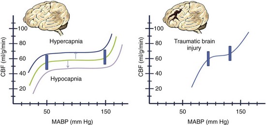
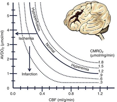


An important implication is that if brain dysfunction is occurring clinically because compensatory mechanisms (e.g., autoregulation changing the vascular resistance, capacity to elevate mean systemic arterial pressure, ability to increase O2 extraction) have been exceeded, the tolerance for further decline in blood flow is low, and tissue damage is seriously threatened. Therapy to increase blood pressure or decrease ICP may be urgently needed. When time permits because the dysfunction fluctuates chronically, it may sometimes be appropriate to measure O2 extraction ratios as one index of the overall adequacy of the CBF. At a low CBF, O2 extraction is increased with a lower venous PO2. It is interesting to note that the variations in CBF and extraction ratios related to neuronal activity are said to underlie the ability to image function by functional magnetic resonance imaging (MRI), a technique that is finding wider usage in the clinical neurosciences.


A fourth principle derives from the other three and the fact that injured tissue swells, making obvious the potential for a cascading injury by a vicious cycle mechanism (Fig. 68-3). If the stage of compensation (see earlier), even with therapy, is exceeded, and ICP is elevated high enough by some mechanism so that cerebral perfusion pressure (CPP) declines, CBF can decline to levels at which tissue injury occurs.
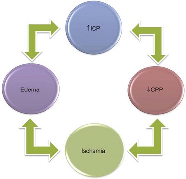


image


Brain edema swelling within the closed cranium will lead to further increases in ICP with even further decreases in CPP in a stage of decompensation. When the capacity for autoregulation is exceeded or damaged so that it can no longer play a role, CBF is linked directly to the CPP.


In the management of intracranial pathology, ICP and CPP are easy to measure continuously and thus serve as highly practical surrogates for the more fundamental, but much more difficult to measure, CBF. However, these are not equivalent, and the limitations of these parameters for guiding therapy need to be remembered. Regardless of causation, when concern arises about the possibility of cascading injury, every effort is made to keep the CPP in the realm of 60 mm Hg (range, 50 to 70 mm Hg) and ICP below 20 mm, Hg if possible. Routinely using pressors and volume expansion to maintain CPP higher than 70 mm Hg is not supported based on systemic complications.3


A fifth principle concerns focal mass effect and its progression in regard to the complex anatomy of the cranial cavity. The cranial cavity is not just a hollow spherical space but contains several almost knifelike projections of folded dura, the falx and tentorium, which divide the cavity into right and left supratentorial compartment and an infratentorial compartment, the posterior fossa. The sphenoid wing is a prominent, mostly bony ridge that separates the anterior fossa containing the frontal lobe from the middle fossa containing the temporal lobe. A narrow opening, the incisura, edged by the tentorium, surrounds the midbrain and is the only passage between the supratentorial and infratentorial compartments. Apart from the small openings for the cranial nerves and arteries, the foramen magnum is the only sizable opening from the cranial cavity as a whole.


The condition that classically illustrates the expanding mass lesion is the acute epidural hematoma, seen after trauma with skull fracture. Regardless of the source, however, the progression can be similar and has been termed rostrocaudal decay to reflect the early and late stages, as listed in order below:


At stages beyond tentorial herniation, it is unusual for focal mass effects not to be accompanied by an overall increase in ICP. The point at which focal mass effect evolves to include a rise in overall ICP depends largely on the compliance within the cranial cavity. Young patients with so-called tight brains can develop raised ICP, even with relatively small volumes of mass that produce only effacement of the cortical gyri. On the other hand, patients with advanced cerebral atrophy can, for example, tolerate large frontal intracerebral hematomas or chronic subdural hematomas with compression of the lateral ventricle and midline shift while maintaining a tolerable ICP and a surprising degree of intact neurologic function.


The Glasgow Coma Score (GCS; see later, “Traumatic Brain Injury”) provides a clinical functional measure of the degree of mass effect and advanced raised ICP. Although it is a useful standardized functional measure of the more advanced stages of mass effect or raised ICP, it was never really intended to focus on more subtle functional changes. A recently introduced coma scale by Widjicks and colleagues4 has delineated the FOUR score (full outline of unresponsiveness) consisting of four components (eye, motor, brainstem, respiration). Each has a maximal score of 4 and takes into account the subtleties related to brainstem activity and breathing patterns occurring during the rostrocaudal decay described.


A sixth principle concerns the separateness of the phenomenology of the following: (1) focal mass effect (as described earlier); (2) diffuse raised ICP; and (3) ventriculomegaly (enlargement of the cerebral ventricles). Although these three processes often occur in combination, the notion that they are separable comes from the observation that there is a pure form of each. The ability to recognize these separate phenomena clinically is often useful in deciding priorities for diagnosis and treatment. This requires some explanation. The pure form of raised ICP—without focal mass lesion, trauma or enlargement of the ventricular system—is a condition known as idiopathic intracranial hypertension or pseudotumor cerebri. The pure form of ventriculomegaly is a condition known as adult chronic idiopathic or normal-pressure hydrocephalus (NPH). The pure form of the focal mass lesion without increased ICP or ventriculomegaly is common; it occurs with tumors too small to raise ICP on their own that are not in a location to interfere with CSF pathways. Instead, there focal mass lesions are typically discovered incidentally or because of symptoms from a focal neurologic deficit or seizure disorder.


Normal ICP varies over a wide range, with generally accepted values between 0 and 20 mm Hg. Diffuse raised ICP, in the fully evolved pure form, results in a clinical picture that may include symptoms of headache, nausea and vomiting, double vision, and obscuration of vision. The accompanying clinical signs may include papilledema and sixth cranial nerve palsy with lateral rectus weakness and side by side diplopia, initially worse on far vision or gaze directed toward the side of the palsy. The papilledema is a mostly chronic phenomenon and is not seen acutely. The sixth nerve palsy of raised ICP can occur regardless of its cause and does not imply direct involvement by a mass lesion, large or small, on the sixth nerve. In this situation, the sixth nerve palsy is a false localizing sign. With raised ICP, there may also be obscurations of vision, in which patients report that their vision temporarily fades or becomes gray, in combination with headache. Again, these obscurations are caused by the effect of diffusely increased ICP on the sensitive optic nerves. They do not imply the presence of a focal mass lesion directly affecting the optic nerves or pathways. Intuitively, it seems that if there is a slow increase in a process raising ICP, the pressure would also rise slowly and evenly, in pace with the evolving process. However, as first shown by Lundberg in 1960,5 the intermediate stages of decompensation are characterized by transient pronounced elevations in ICP (to 60 mm Hg) that characteristically plateau for up to 45 minutes and then transiently cycle down again to a more normal range.


The original form of this condition of chronic, diffuse, raised ICP is known by the old term pseudotumor cerebri or the more descriptive idiopathic intracranial hypertension. Because it is often not benign, causing disabling chronic headache and visual loss, sometimes even to permanent blindness, the term benign intracranial hypertension is falling out of favor. The basis for the condition is not entirely understood. It usually occurs in obese young women. Treatment is with acetazolamide diuretics, steroids, and intermittent lumbar puncture. Severe cases with threatened permanent visual loss may require a lumboperitoneal or ventriculoperitoneal CSF shunt or optic nerve fenestration, in which the meninges around the optic nerves are opened to vent CSF in the orbit.


Pure ventriculomegaly—specifically, enlargement of the lateral ventricles—is characterized by gait disturbance and incontinence early in the clinical picture. As the process worsens, cognitive disturbances may be added on. The early appearance of gait disturbance and urinary incontinence is attributed to dysfunction of the medial cerebral hemispheres in which the leg area of the primary motor cortex and bladder control area reside. Nerve fiber pathways inside the brain must pass around the lateral ventricles to reach these areas on the medial hemisphere and therefore are especially vulnerable to pressure or distortion by the enlarged ventricle. This syndrome is called NPH. The usual diagnostic difficulty is in differentiating the condition from cerebral atrophy. Ventricular enlargement more prominent than enlargement of the CSF subarachnoid spaces over the cerebral convexity is typically seen in NPH. The clinical impression that gait disturbance and incontinence occur early and predominate over dementia is considered an important feature of NPH. Treatment is with CSF shunt, lumboperitoneal or ventriculoperitoneal. The differentiation between NPH and cerebral atrophy is important because of the increased risk for subdural hematoma with shunting in cerebral atrophy.


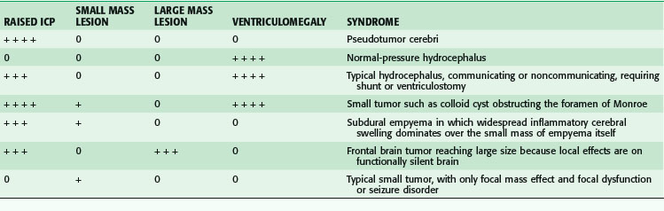
Table 68-2 summarizes the relationships among ICP, mass lesions, and ventriculomegaly.


Cerebrovascular Disorders


Cerebrovascular disorders encompass a host of disorders, congenital and acquired (Box 68-1).


Arteriovenous Malformation and Fistula


In the early embryonic stage, the circulatory system does not yet have capillaries between the arterial and venous sides. Instead, there are vascular channels approximately 200 µm in diameter that must undergo further development and maturation to form the capillary bed. There is reason to believe that when this does not occur perfectly, a focal failure of maturation can lead to a nidus of persistent embryonic, low-resistance vessels connecting the arteries and veins. Over time, the high blood flow in these circuits leads to secondary changes that enlarge the nidus and the afferent arteries and efferent veins, often to impressive proportions. The high flow in the nidus, afferent, and efferent vessels predisposes to degenerative events, sometimes with aneurysm formation, causing hemorrhage, intracerebral, subarachnoid, or both. Brain tissue at the edge or intermixed with the abnormal vessels may develop dysfunction to become an epileptic focus or, less commonly, progressive ischemic deficits occur as the low-resistance AVM draws blood flow away from adjacent areas with normal vascular resistance.


It follows that AVMs can have a wide variety of configurations and sizes, depending on which part(s) of the vascular bed fail to mature and which consequences of the increased flow occur over time. If the venous outflow is restricted, the venous side of the complex may enlarge disproportionately and form a venous varix; so-called vein of Galen aneurysm is the prime example. Here, the vein of Galen, restricted by the downstream outflow limitations of the stiff dura–contained straight sinus, dilates, sometimes massively, and can cause obstructive hydrocephalus in the newborn, often together with high-output heart failure. Usually, the venous outflow channels enlarge to a moderate degree and become thickened in the vessel wall, or arterialized. They do not generally cause symptoms by mass effect.


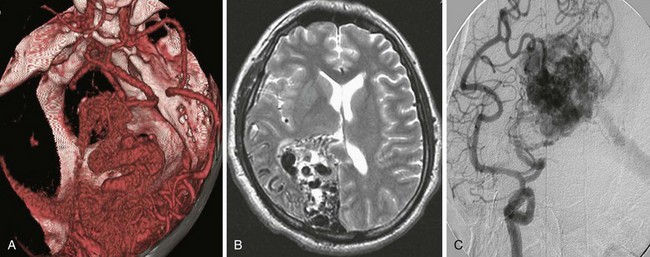
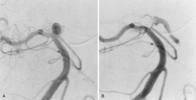
Clinical presentation is usually that of a hemorrhagic stroke picture, typically an intracerebral or subarachnoid hemorrhage, or some combination. The patient complains of sudden-onset severe headache with or without focal neurologic deficit and meningismus. These symptoms can occur in all degrees of severity, but are less commonly fatal than after aneurysmal subarachnoid hemorrhage into the basal cisterns. Investigation usually begins in the emergency department with a computed tomography (CT) scan showing the hemorrhage. MRI or magnetic resonance angiography (MRA) studies typically follow, often showing enlarged afferent and efferent vessels. The role of CT angiography is evolving. Regardless of the mode of presentation, diagnosis is ultimately made by conventional catheter cerebral angiography (Fig. 68-4). It is based on demonstration of arteries and veins on the same conventional angiographic image, proving the high-flow shunting of blood through the nidus network or fistulous vessels. In an arteriovenous fistula, the shunting occurs through short, sparse, larger-diameter channels so that a cloudlike nidus of smaller vessels is not evident. Instead, the enlarged afferent arteries appear to connect directly with the enlarged efferent veins. In the typical AVM, there is a cloudlike nidus, or network of smaller vessels, seen well on angiography but not necessarily fully appreciated on MRI or MRA. The AVM can occur in all locations and with varying degrees of size, complexity, and compactness.


When the patient presents with a new-onset seizure, the next most common presentation, the investigation typically begins with brain MRI showing enlarged afferent and efferent vessels. MRI, however, may not suggest the diagnosis if the afferent and efferent vessels are not grossly enlarged; however, this is rare in lesions presenting with seizure. Again, definitive diagnosis is with conventional catheter angiography, but this may sometimes be omitted if the diagnosis is sufficiently suggestive on MRI alone and there is reluctance to consider surgery, with only anticonvulsant treatment planned.


It is important to pay attention to the nidus or fistula as the prime mover of the process and realize that the secondary changes in the afferent and efferent vessels, however impressive they may seem, will generally revert to normal once the nidus or fistula has been resected or occluded. The assessment of AVM size refers to the size of the nidus and not the conglomeration of large feeding or draining vessels.


Therapy can include surgical excision, endovascular embolization of the nidus, and/or SRS. The decision to embark on any treatment depends on the assessment of the patient’s treatment risk in comparison with the natural risk. The studies of Ondra and associates6 have fairly well characterized the natural risk. Hemorrhage occurs with a frequency of approximately 4%/year, and because only approximately 25% of these are severe, with permanently severe disability or death, the catastrophe risk is only approximately1%/year. The treatment risk, therefore, must be favorably low to justify action in most situations, and delays in treatment to allow for patient acceptance and optimize the circumstances for therapy are understandable and reasonable. The main reason to recommend treatment is that the treatment may offer a lower risk over the long term. Younger patients have the most to gain by such an assessment. It may be possible to define features of individual AVMs that adjust the natural risk upward or downward, but so far these have been difficult to prove. For the purposes of clinical decision making, the hemorrhage risk is generally taken across the board for all AVMs as a starting point. An across-the-board assessment of treatment risk, however, is clearly not warranted because each AVM. Size, location relative to access by the treatment method considered, and location relative to proximity to deficit-prone brain structures are all important variables to consider. Another feature is the compactness of the AVM, with some forming a tightly clustered nidus, with little brain tissue in between, and others being diffuse and rambling, with scattered small clusters of nidus vessels encompassing large intervening areas of functional brain.


Obviously, the smaller, more compact AVMs located superficially in areas of silent brain function are the most attractive for open surgical resection. Diffuse large AVMs encompassing deficit-prone areas of brain are least attractive to open surgery and other methods. Spetzler and Martin7 have devised a grading system for assessing risk with open operation. However, any decision is ultimately based on how an individual neurosurgeon assesses the risk for that AVM in a patient, with his or her limits of risk tolerance.


Surgical excision is carried out by craniotomy and microsurgical techniques. The lesions are evident at operation as a bright red blood–containing network of vessels. Feeding arteries are thicker walled than the generally larger, thinner walled draining veins. Nidus vessels are thin, bright red, and very thin walled, so that they resist coagulation. This feature makes it important to remain just at the edge of the lesion, if possible. Entry into the AVM nidus results in vigorous bleeding, which is time-consuming to control. Recent innovations in surgical therapy include the more common use of frameless stereotaxy or neuronavigation, which allows more accurate localization and definition of the margins, important feeders, and draining veins. Also, newer bipolar cautery instruments with advanced nonstick features have been helpful. A risk of open surgery is the unintended occlusion of vessels passing through the lesion to supply important functional areas of brain. Also, there is the potential to leave part of the nidus behind inadvertently, so the hemorrhage risk would be unpredictable. Postprocedure angiography is essential to perform at the end of the procedure, or as soon as possible thereafter. Any residual AVM is addressed as soon as practical. Another category of concern is hemorrhage resulting from a sudden increased blood flow at the periphery of the resected lesion, exceeding the limited autoregulatory capacity there. For the larger more complex AVM, intentionally staged surgery is sometimes an option and is probably reasonably safe as long as the remaining venous drainage of the AVM stays in balance with its residual afferent blood supply at each stage.


SRS is most attractive for small (<2.5 cm diameter) deep lesions that are difficult to access by open surgery. This method has the obvious attraction that hospitalization and craniotomy can be avoided, and patient acceptance is often high. The size of the nidus is the major limitation, however; that the smaller the nidus, the higher the dose of radiation that can be given safely and the more effective the treatment is likely to be. Negative considerations are that the radiation is not immediately effective and the risk for hemorrhage during the latent interval must be factored in. Approximately 60% of treated AVMs will be obliterated in 1 year (and ≈80% by 2 years). The obliteration rate is probably higher in younger patients and in those with nidus vessels of smaller diameter. The larger normal vessels generally escape obliteration by SRS, even when passing through the treatment volume to supply distant areas of the brain. However, this may also mean that the more fistulous type of AVMs, with sparse, large-diameter nidus channels, is less likely to be treated successfully. When an AVM reacts to SRS, there may be a period of edema in the surrounding or intervening brain. Usually, the edema evident by MRI is not symptomatic. However, the edema can be extensive and sometimes is temporarily disabling for up to several months. Steroids may be helpful in this setting. Seizures may also be more likely during a reactive interval and require upward adjustments of anticonvulsant medication, but the eventual seizure risk is not increased. Overall risk for permanent neurologic deficit with SRS is low, approximately 2% or 3%, when pooling large numbers of patients but, as with other treatment modalities, the risk for permanent deficit is dependent on proximity to deficit-prone structures. The optic nerves and chiasm and brainstem are of particular concern with radiation. It is safest not to target lesions within 5 mm of the optic pathways, which limits its application in treating parasellar or pituitary lesions.


Endovascular therapy is a rapidly evolving field, making it difficult to assess in terms of predicting actual patient outcomes with methods currently available. Whatever criticisms apply currently may be overcome with newer methods, but that remains to be proved. It is increasingly being carried out by neurosurgeons, although the familiarity with catheter angiography has made it largely the domain of the interventional neuroradiologist. Endovascular therapy has the allure of avoiding open surgery, but the likelihood of obliterating an entire AVM by this method alone is limited at present. To be effective in any permanent way, the embolic material must reach the nidus vessels but not pass readily through to the venous side. This can be more difficult than it seems because the diameter of the nidus vessels may not be uniform. Initially, the high blood flow to the nidus draws the embolic material to it but, as the nidus progressively occludes, the embolic material is progressively more likely to reflux into normal vessels and the risk for stroke increases. The method is safest in situations in which there is a long segment of feeding vessel dedicated to the AVM because then reflux to the normal circulation is less likely. As with any endovascular technique, some areas of the circulation are easier to access than others. The method can be effective alone for treating the smaller, more fistulous lesions with single feeding and draining vessels. These lesions are rare, however, and the method is generally used to reduce the size or complexity of an AVM just before open surgical resection. However, afferent vessel occlusion alone, without penetration of the nidus, is not effective in any other context. Over time, an occluded feeding vessel recruits a network of replacement channels that may make it even more difficult to treat definitively. When used in conjunction with radiosurgery, the embolic material must reach the nidus, reducing the nidus volume permanently in a dimension and making radiosurgery of the remainder more effective. Risks for permanent deficit with embolization have been described as high as 5% per embolization session, but this figure is based on older techniques. The problem with clinical decision making is that currently we do not know the risks of evolving techniques accurately.


Cavernous Angioma: Cavernous Malformation


The cavernous angioma is a highly characteristic, usually almost spherical, discrete lesion, composed of a cluster of vascular sinusoids fed by small vessels in the arteriolar size range or smaller. Unlike the AVM, large feeding arteries and draining veins are not seen, and diagnosis by catheter angiography is not possible. The sinusoids are tightly compacted and there is no intervening brain tissue, a diagnostic feature. At surgery, they often appear as a discrete, mulberry-like collection of thin-walled vascular sinusoids with a greenish hemosiderin rim in the surrounding brain edge. When incised, bleeding from the lesion is minimal and easy to control, unlike the AVM, which bleeds vigorously if entered. The sinusoids often seem to be admixed with small chronic hematomas containing blood in various stages of decomposition. Many are older and contain birefringent yellow cholesterol crystals. Early autopsy studies8 noted out that they should be more common than recognized clinically. However, the lesions were not usually evident on CT or conventional catheter angiography available at that time. The clinical entity was not fully appreciated until the more widespread availability of MRI in the 1980s. The lesions are easily seen on MRI, where their appearance is diagnostic. They show a center containing some small, high-intensity signal foci surrounded by a null signal corona, dark on T1- and T2-weighted images, that corresponds to hemosiderin deposition in the adjacent brain.


Like the true AVM, cavernous angiomas can present with symptomatic hemorrhage or new-onset seizures. Unlike AVM, they can also present as a slowly growing mass lesion. At least part of this growth appears to result from the accumulation of multiple small hemorrhagic foci in various stages of healing. The lesion can be particularly problematic in this manner when located in the brainstem. The hemorrhage frequency is not understood completely, partly because most hemorrhages are probably small and asymptomatic and not recorded as events. When located in the cerebral cortex or medial temporal lobe structures, they can be a focus of seizures and can be solitary or multiple, incidental or symptomatic. The multiple form is more common in women and in those of Hispanic descent and has been linked to the KRIT-1/CCM1 gene locus on chromosome 7q21.2. Cavernous angioma can appear de novo in patients with earlier, completely negative MRI scans. Because of these features, cavernous angiomas occupy a position somewhere between congenital vascular malformations and vascular tumors. The term angioma probably reflects this tumor-like nature better than the term malformation and is preferred here for that reason.


Many lesions are followed conservatively with interval MRI scans for years, without signs of activity. At the other extreme, they can sometimes bleed to produce a large symptomatic or life-threatening intracerebral or brainstem hematoma, but this is relatively uncommon. The threat of such hemorrhage is generally not held out as a reason to operate, as it is for the true AVM. Surgical excision is usually reserved for lesions that are problematic because of hemorrhage, demonstrated growth, or seizures difficult to control with medication alone. Surgical excision is practical in most locations because of the distinct margins and minor bleeding encountered at operation. Lesions in the brainstem can pose major challenges but, even there, surgical resection is not ruled out as long as access can be achieved without the need to traverse crucial structures anticipated to produce a major new deficit. Intraoperative ultrasound and frame-based or frameless stereotaxy can be helpful in locating the lesions if they are small and in deep locations. Radiosurgery can be performed for these lesions but is not of demonstrated benefit. The hazards of radiosurgery in critical brainstem locations remain a concern.


Traumatic Arteriovenous Fistula


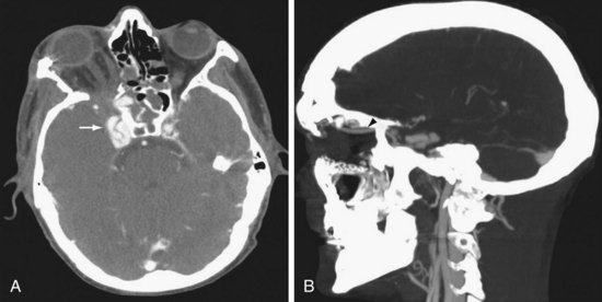
Both the internal carotid artery (ICA) and vertebral artery enter the cranial cavity immediately after passing through a venous network. The ICA passes through the cavernous sinus, which communicates with the superior ophthalmic vein, petrosal sinus, and sphenoparietal sinus. The vertebral artery passes through a venous plexus at the occipital-C1 epidural space, which communicates with the jugular vein, epidural venous plexus, and paraspinal venous plexus. Trauma leading to a tear in the carotid or vertebral artery at its tether point passing through the skull base can lead to fistula with the surrounding venous plexus. The consequences may vary in severity and suddenness but typically include periorbital swelling, with proptosis and scleral edema in the case of the carotid-cavernous fistula (CCF) and prominent pulsatile bruit in the case of the vertebral-jugular fistula. Intraocular pressure measurement by tonometry can guide the urgency in treating CCF. Radiologically, dilation of the superior ophthalmic vein is characteristic (Fig. 68-5). These lesions are usually treated by endovascular techniques. A catheter is advanced through the tear in the artery into the venous side of the fistula. The high-flow and large fistulous channel facilitate this process. Embolic material, a coil, or a detachable balloon is then used to occlude the venous side of the fistula. When conventional transvenous routes fail, a direct approach via transorbital puncture may be required to provide endovascular therapy.9


Cerebral Saccular (Berry) Aneurysms


Saccular or berry cerebral aneurysms form as a degenerative change in the wall of the larger intracranial arteries in and around the circle of Willis. They are rare in children and occur with increased frequency in older age groups, sooner in patients with connective tissue defects, such as polycystic kidney disease, Marfan syndrome, and Ehlers-Danlos syndrome.


Aneurysms form in relation to defects in the smooth muscle–containing media layer. Such defects are common at branch points in the arteries and can also occur in relation to shear forces at the edge of stiffened parts of the vessel wall containing atheroma. The defect in the media then allows the intima to stretch outward, fragmenting the internal elastic lamina in the process and carrying the connective tissue of the external adventitial layer outward with it. The connective tissue in the dilating intima and adventitia is capable of proliferation so that the aneurysm can reach a size considerably larger than stretching alone would allow. The Laplace equation predicts that at any given pressure, the stretching force on the wall of the aneurysm increases as the diameter increases. The process is therefore inherently not self-limited, but progressive. With robust connective tissue proliferation in the wall, a minority of aneurysms enlarge to surprising dimensions; hence, the blood flow within slows to allow thrombus formation, usually in concentric layers, to form a partly solid mass of much greater outer diameter than the lumen would suggest on angiography alone.


Distal embolization of clot material is a rare occurrence. Calcification can occur in the wall in advanced cases and the adjacent brain can become gliotic from chronic pressure, making seizures also a possible presentation. An enlarging aneurysm can also compress an adjacent cranial nerve; the optic and third nerves are usually affected by this mechanism. Generally, however, it is much more common for an aneurysm to present with subarachnoid hemorrhage. The proliferative ability of the connective tissue in the dome of the forming aneurysm can be exceeded by the stretching force, leading to rupture. The onset is unpredictable and appears to occur at a surprisingly low rate. Incidentally discovered unruptured aneurysms bleed at a rate depending on size. Those smaller than 1 cm bleed at a rate of 0.05% to 0.5%/year, whereas those larger than 1 cm bleed at a rate of 1% to 2%/year. Treatment recommendations for unruptured aneurysms smaller than 5 mm in diameter vary widely based on patient preference, aneurysm accessibility to treatment, and surgeon’s assessment of risk. Once hemorrhage from an aneurysm occurs, the situation changes dramatically. Bleeding of highly oxygenated arterial blood occurs suddenly into the surrounding CSF-containing subarachnoid space, which initially offers little backpressure. Aneurysmal subarachnoid hemorrhage can occur in all gradations of severity. In most patients, accumulated blood in the basal spinal fluid cisterns leads to a coagulum that spontaneously stops the bleeding. In 10% to 15% of patients, the bleeding is so severe at the outset that death occurs before they even reach the hospital. Approximately 40% die following the initial hemorrhage but at a later stage. Rebleeding occurs with a peak incidence in the first 24 hours after the initial event. If the aneurysm is left unsecured, the rebleed rate is 20% in the first 2 weeks, 50% in the first 6 months, and thereafter 3% to 4%/year.10 Rebleeding is the principal cause of death, usually by raised ICP.


Subarachnoid hemorrhage, with or without increased ICP, sets in motion a series of problems that can cause complications and poor outcomes, regardless of the technical success of treating the aneurysm itself. First, the blood can interfere with the spinal fluid circulation so that acute hydrocephalus can result in a raised ICP and reduced cerebral perfusion. The hydrocephalus is evident as ventricular enlargement on CT and can usually be readily treated with ventricular drainage. Second, the highly oxygenated blood coagulum surrounds the vessels traversing the subarachnoid space. Whereas these vessels are usually in an environment of clear colorless CSF, they now are in a milieu of decomposing blood, triggering the activation of lysosomal and proteolytic enzymes and generating chemically active free radicals. The smooth muscle coating of the otherwise intact vessels can become irritated to trigger vasospasm, at first reversible but, in severe cases evolving to a damaged and swollen arterial wall, with persistent luminal narrowing. The vessels eventually remodel over 3 to 6 weeks and return to a normal configuration but, all too often, ischemic deficits in the supplied brain occur in the interim. Unfortunately, these ischemic neurologic deficits are not rare and are the single major cause of serious morbidity after successful aneurysm treatment following subarachnoid hemorrhage. The blood coagulum eventually clears from the subarachnoid space but sometimes initiates a progressive slow fibrosis that results in delayed hydrocephalus, distinct from the early hydrocephalus caused by the coagulum itself. Cerebral vasospasm following subarachnoid hemorrhage starts on days 3 to 5 and peaks during days 5 to 14, resolving slowly over the weeks following the initial insult.


Treatment of the vasospasm includes elevating the blood pressure, blood volume, and cardiac output in an attempt to bring more blood flow past the narrowing in the vessels (Box 68-2). The calcium channel blocker nimodipine appears to reduce the incidence of delayed ischemic neurologic deficits and prevents vasospasm, probably through opening collaterals to the ischemic brain. Its direct effect on the vasospasm is still in question. Phase IIA trials with clazosentan, an endothelin A antagonist, have been promising in reducing the frequency and severity of cerebral vasospasm.11 Attenuation of vasospasm by statins or magnesium sulfate is controversial according to recent literature.12,13


Investigation of patients with a suspected aneurysmal subarachnoid hemorrhage begins with an assessment of the history. Because the hemorrhage occurs into the subarachnoid space and not brain tissue, there is usually no focal neurologic deficit. Symptoms of sudden-onset headache with meningismus are classic and reflect a sudden rise in ICP and irritation of the basal meninges by the blood. In severe cases, the patient may be comatose or uncooperative. A report of a strokelike picture of sudden onset of neurologic signs and symptoms should prompt CT scanning, which reveals blood filling the subarachnoid cisterns (Fig. 68-6A). Because the hemorrhage can occur in all gradations of severity, the difficulty comes in recognizing the patient with the small sentinel hemorrhage who arrives in good condition with only the complaint of an alarmingly severe sudden onset of headache. Headache from increased ICP may be only transient until compensatory mechanisms occur. Residual headache and neck stiffness from meningismus, although usually present, may not be impressive.


A high index of suspicion is required for these patients. It is important to focus on the suddenness of symptom onset rather than severity because reports of severity are more subjective or may be tainted by a strong psychological overlay of denial or exaggeration; also, the headache caused by a very small hemorrhage may be dissipating naturally to some extent by the time the patient is finally assessed. If the headache onset is truly or almost instantaneous, lumbar puncture is usually advised in cases in which the CT scan is negative. Blood in the CSF with xanthochromia in the supernatant is diagnostic.


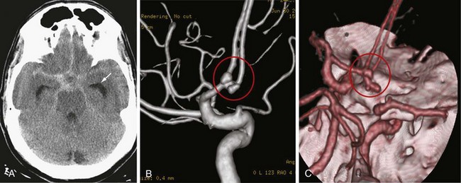
If the CT scan or lumbar puncture is positive for subarachnoid blood, the next step is usually conventional catheter cerebral angiography (see Fig. 68-6B). CT angiography with bolus IV contrast has become increasingly convenient and accurate; thus, it is being used more frequently. Newer scanners are capable of large numbers of simultaneous slices so that three-dimensional reconstruction with good registration and surprising detail is possible (see Fig. 68-6C). With selections in density windowing, the relation of the aneurysm to any blood clot can also be visualized.


Treatment of a cerebral aneurysm can be performed in a variety of ways:


Segmental occlusion and bypass are usually reserved for those cases that are awkward to treat by any other method. The choice of endovascular coil occlusion versus open surgical clipping has, on the other hand, become a complex issue.


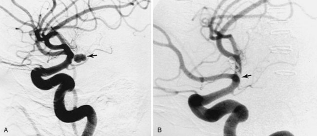
In terms of the aneurysm configuration, a narrow neck is a favorable attribute for treatment by open surgical clipping or endovascular embolization methods. Wide-necked or sessile aneurysms with a high neck-to-fundus ratio make it difficult to contain coils in the fundus without protrusion into the parent artery. A stent placed in the parent artery may address this problem, but the technology is evolving and stenting is currently practical only in more proximal locations. Generally, wide-necked aneurysms gravitate toward open surgical treatment, although stent-assisted coiling can be also used in some cases. Particularly interesting is the recently introduced concept of functional reconstruction with the use of flow diverters. These stents induce reverse remodeling and delayed disappearance of the aneurysm via thrombosis. They are especially useful for very small, giant, wide-necked, or otherwise difficult to treat aneurysms and can be used with intramural coil placement.14,15


In regard to aneurysm location, the more proximal the aneurysm, the easier it is to reach by endovascular means, whereas the more distal the aneurysm, the more difficult it is to reach and treat effectively by that method. Aneurysms in the cavernous sinus are considered difficult to reach by any open surgical method and are generally only treated by endovascular means or by parent artery occlusion with bypass. Aneurysms of the ICA at the level of the ophthalmic artery origin can be treated by open surgery or endovascular methods, as can most aneurysms of the ICA, proximal middle cerebral artery (MCA), and anterior cerebral artery (ACA), as far as the anterior communicating artery. The more distal MCA aneurysms, at the trifurcation in the sylvian fissure and beyond, and ACA aneurysms distal to the anterior communicating artery are more difficult to treat by endovascular methods and generally are treated by open surgical clipping. The basilar artery termination and midbasilar area can usually be reached easily by endovascular methods, whereas open surgery is possible but not feasible in these locations. The vertebral artery aneurysms can be reached easily by either method.


In situations in which the aneurysm location or configuration presents a clear advantage of one method over the other, decision making is fairly easy, even at centers at which all current methods are available. Problems in decision making involve aneurysms that are favorably situated and configured so that they would be attractive to treat by open surgical clipping or endovascular coil occlusion. In the past, open surgical clipping was considered first,and the patient was referred for endovascular coil occlusion only if there were reasons to avoid open surgery. However, since the European prospective randomized trial of surgery versus open clipping, this paradigm is now open to question; it is becoming more common to consider endovascular treatment before open surgical clipping in aneurysms treatable by either method.16


Increasingly, the patient is treated by a team approach in which all treatment methods are available and considered in view of the patient’s aneurysm location, configuration, general medical condition, and expressed personal or family preference(s). Endovascular treatment avoids craniotomy, but has an aneurysm recurrence rate of 15% or higher. The recurrence rate after open surgical clipping appears to be much lower.


Results of treatment of unruptured aneurysms are generally good by any method, but these usually involve a carefully selected group for whom treatment conditions are favorable and, most importantly, there is no aftermath of subarachnoid hemorrhage to overcome. These beneficial results in patients with unruptured aneurysms, without subarachnoid hemorrhage, contrast markedly with the overall disappointing outcomes in patients with aneurysmal subarachnoid hemorrhage. Regardless of the type of treatment, the risks for disabling ischemic complications from cerebral vasospasm and hydrocephalus are much higher in patients with subarachnoid hemorrhage. Patients with aneurysmal subarachnoid hemorrhage who present at the hospital in reasonable condition, at least awake and talking, still have only a 60% chance of returning home without functional deficit. The treatment contributes approximately 5% to the complications. Vasospasm and the comorbidities associated with prolonged treatment account for most of the rest. Infection from the ventriculostomy needed to treat hydrocephalus and control ICP is a minor component of the complication rate.


Spontaneous Intracerebral Hemorrhage


Spontaneous intracerebral hemorrhages into the brain parenchyma are common, accounting for approximately 10% of all strokes. They generally occur in older patients, usually because of degenerative changes in the cerebral vessels that are often associated with chronic hypertension (Box 68-3). In younger patients, they are more likely related to drug abuse or vascular malformation. They can occur anywhere in the cerebral circulation or brainstem but are classically described in association with small degenerative aneurysms (microaneurysms; also known as Charcot-Bouchard aneurysms) at the junctions of the perforating vessels and larger vessels at the skull base. They are typically on the MCA junctions with the small perforating lenticulostriate vessels, leading to hemorrhage into the putamen. The clinical presentation is with a stroke pattern of sudden-onset neurologic signs and symptoms that depend on the area of brain affected. Symptoms are more likely to include headache than ischemic stroke. The diagnosis is with CT, usually done in an emergency department setting. The size and location of the acute hematoma are well visualized with CT, as well as any associated brain shift or hydrocephalus (Figs. 68-9A and 68-10). In older patients with a known history of hypertension and classic CT appearance of a hematoma in the putamen, thalamus, cerebellum, or pons, further diagnostic studies are generally not indicated. Rehemorrhage is unlikely in that setting. However, further investigation might be warranted with an atypical hematoma location or appearance, especially if there is any component of subarachnoid blood. Also, investigation is usually recommended for younger patients without known hypertension and those with a potential underlying cause for hemorrhage (e.g., history of neoplasm, blood dyscrasias, bacterial endocarditis).


Further investigation is generally done with contrast MRI or MRA. Any suggestion of aneurysm or AVM is followed by conventional catheter angiography. In older patients with a history of early dementia and multiple episodes of more peripherally located intracerebral hematomas, the diagnosis of amyloid angiopathy needs to be considered.


Most cases of spontaneous intracerebral hemorrhage do not require an operative procedure. Many hemorrhages are small enough to be well tolerated and do not require surgery. Others are so large at the outset that surgery is of little benefit. Relief of any obstructive hydrocephalus by ventricular drainage is usually offered, except in the most impossible cases. Patients who obey commands and can be monitored by changes in their neurologic examination can generally be managed conservatively with hospital observation for at least 5 to 7 days. Peak swelling and decompensation are probably most likely to occur within that time frame. Surgery for evacuation of the hematoma may be appropriate in a small group of patients with intermediate-sized hemorrhages in accessible locations who appear to tolerate the hematoma initially, but then deteriorate in a delayed fashion with edema, despite medical therapy. Steroids have not demonstrated benefit. Attempts to predict which patients will deteriorate based solely on hematoma volume have been frustrated by the broad spectrum of intracranial compliance exhibited by different patients. Generally, younger patients with smaller ventricles and small subarachnoid spaces have a lower compliance, with lower tolerance, than older patients with cerebral atrophy and generous ventricles and subarachnoid spaces.


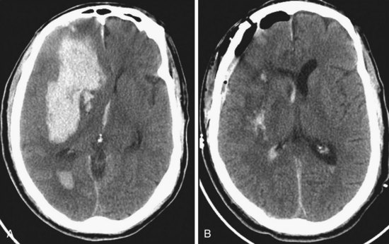
The Surgical Trial in Intracerebral Hemorrhage has noted a lack of clinical outcome difference when comparing early surgery with conservative management.17 If indicated, surgical evacuation is usually done by craniotomy over the most accessible part of the hematoma (see Fig. 68-9B). Intraoperative ultrasound is often helpful in finding hematomas that do not quite come to the cortical surface and in monitoring the progress of the evacuation. The goal of surgery is decompression more than complete removal, but is generally done as far as safely practical. The wall of the hematoma cavity is inspected for any underlying cause and a biopsy is taken, if indicated. Putamen hemorrhage can sometimes be evacuated with minimal surgical damage to the overlying brain by a trans-sylvian fissure–transinsular approach. Stereotactic aspiration and methods with fibrinolytic agents are being developed and may be a consideration for patients with hematomas in deep locations that are otherwise difficult to access.


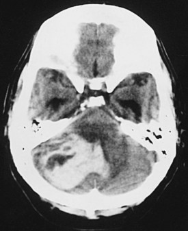
A special situation to consider is the patient with cerebellar hemorrhage (see Fig. 68-10). Surgery is offered more readily in these cases because the danger of sudden deterioration from brainstem compression is more of a concern, and because even extensive damage to the cerebellum itself is generally survivable, with good functional outcome. Patients with fourth ventricular obstruction and hydrocephalus from cerebellar hemorrhage can sometimes be treated with ventricular drainage alone but are usually offered surgical evacuation of the hematoma by suboccipital craniotomy because of the risk for brainstem compression.


Mycotic Aneurysms


These aneurysms are associated with a systemic infection capable of showering small particles of bacteria-infected material into the cerebral vascular bed. Subacute bacterial endocarditis and some pulmonary infections can do this. A distinguishing feature of these aneurysms is that they are generally found more distal in the cerebral vascular bed, as opposed to berry aneurysms, which are usually found on larger vessels near the circle of Willis. There can also be many of them. When the bacterial emboli lodge in distal cerebral arterial branches, they can erode through the wall of these smaller vessels, often creating a hemorrhage contained by the perivascular tissue. Maximal antibiotic treatment is essential at the outset. The presence of an intracerebral hematoma may force immediate craniotomy for evacuation. Operation on the aneurysm at this early stage often reveals a component of subarachnoid hemorrhage and an early inflammatory reaction in the subarachnoid space, with only a blood collection covering the erosion defect in the wall of the small artery. Attempts to dissect and define a neck are frustrated by a lack of developed fibrous tissues and intraoperative hemorrhage is then common. Typically, the diseased arterial segment must be occluded and resected when operated on in this early stage. The need for arterial bypass to maintain blood flow to critical cerebral areas should be anticipated, but this is not always possible.


If the mycotic aneurysms are discovered or treated at some later stage, a fibrous wall to the aneurysm may have had time to develop, and clipping can then be a possibility. However, the surgeon needs to be forewarned that it may be difficult to find the aneurysm in a distal location, often buried deep in a cerebral sulcus thickened with reactive fibrous scar tissue.


Moyamoya Disease


Moyamoya disease is a cerebrovascular disorder that is characterized by an idiopathic nonatherosclerotic narrowing or occlusion of major intracranial blood vessels with the development of a conspicuous compensatory collateral rete vessel network, which allows continued cerebral perfusion around the occluded or severely narrowed segment. The disorder is usually bilateral, although not necessarily exactly symmetrical. Although generally rare, the disease is more common in persons of Asian ancestry and was first recognized from cases studied with angiography in Japan before the advent of CT and MRI. The term moyamoya comes from the Japanese word for “puff of smoke,” or mist. The actual disease is sometimes confused with the less conspicuous collateral vascular networks seen around severe narrowing of common atherosclerotic origin in persons of Western origin. In the juvenile form, moyamoya typically presents as cognitive decline, with deteriorating school performance and evidence of multiple infarcts. Angiography reveals the ICA, proximal MCA, or proximal ACA with severe narrowing or occlusion and, generally, multiple clusters of fine collateral vessels. In the adult form of moyamoya disease, the rete vessels cause subarachnoid or basal ganglia hemorrhage, the most common presentations. The hemorrhage can usually be treated conservatively. Some form of extracranial to intracranial bypass is generally attempted to take the load off the collateral vascular network. In younger patients, the results are good, with an onlay interposition of the superficial temporal artery sewed into the dura after a strip craniotomy. A feature of the disorder is the vigor with which collaterals form from the onlay transposed vessel. In the adult form, a microvascular anastomosis with the superficial temporal artery or grafted vessel may be preferred.


Dural Arteriovenous Malformations


Dural AVMs (type 2 CCF is a subtype) are not often seen in younger patients. The lesions seem to occur only in adults and are probably acquired lesions that follow a dural sinus thrombosis, usually of the cavernous sinus or sigmoid-transverse sinus junction area. With subsequent healing, the thrombosed segment triggers a neovascular response that evolves to an AVM configuration with fistulous channels that can gradually enlarge. Usually, there is associated stenosis of the affected dural segment, suggesting the earlier thrombosis. The lesions are generally not dangerous unless they cause retrograde venous drainage into the cerebral circulation. The risk for intracranial hemorrhage is then fairly high; it is important at least to separate the dural AVM drainage from the cerebral circulation when that occurs. In the case of transverse-sigmoid sinus dural AVM, the patient usually complains of a bruit and embolization or resection is optional, depending on symptom tolerance. In the case of type 2 CCF, the problem is usually intraocular and intraorbital venous hypertension, with proptosis, chemosis, and sometimes threatened vision. Ocular tonometry can help determine the extent of the threat to vision. Treatment of type 2 CCF involves endovascular embolization of prominent feeders, followed by occlusion of the affected venous dural sinus. As long as the dural AVM drainage is separated from the cerebral circulation, occlusion of the affected venous drainage is safe and curative. The affected stenotic transverse-sigmoid sinus segment can be reached by endovascular techniques through the jugular vein. The cavernous sinus can be reached by the petrosal sinus or, with neurosurgical assistance, through the superior orbital vein.


Central Nervous System Tumors


Clinical Presentation


The clinical manifestations of various brain tumors can be divided into those caused by focal compression and irritation by the tumor itself and those attributed to secondary consequences—namely, increased ICP, peritumoral edema, and hydrocephalus. Usually, symptoms are caused by a combination of these factors.


The clinical presentation does not differ much by tumor histology but rather by rate of growth and location of the tumor. A meningioma peripherally located in a relatively silent area of the brain, with a slow rate of growth, may enlarge to a significant size in a neurologically intact patient because the brain can accommodate to a slowly growing lesion. On the other hand, a small metastatic lesion at the foramen of Monro or in the sensorimotor strip can cause acute hydrocephalus or seizures, respectively.


Headache occurs in 50% to 60% of primary brain tumors and in 35% to 50% of metastatic tumors. It is classically described as being worse in the morning, probably because of hypoventilation during sleep, with consequent elevation of the PCO2 and cerebrovascular dilation. The headache is associated with nausea and vomiting in 40% of patients and may be temporarily relieved by vomiting as a result of hyperventilation. Seizures may be the first symptom of a brain tumor. Patients older than 20 years presenting with a new-onset seizure are aggressively investigated for a brain tumor.


Infratentorial lesions may present with headache, nausea and vomiting, gait disturbance and ataxia, vertigo, cranial nerve deficits leading to diplopia (abducens nerve), facial numbness and pain (trigeminal nerve), unilateral hearing deficit and tinnitus (vestibulocochlear nerve), facial weakness (facial nerve), dysphagia (glossopharyngeal and vagus nerves), and CSF obstruction causing hydrocephalus and papilledema. Supratentorial lesions may present with different symptoms, depending on the location. Frontal lobe lesions manifest as personality changes, dementia, hemiparesis, or dysphasia. Temporal lobe lesions may present with memory changes, auditory or olfactory hallucinations, or contralateral quadrantanopsia. Patients with parietal lobe lesions may develop contralateral motor or sensory impairment, apraxias, and homonymous hemianopsias, whereas those with occipital lobe lesions may show contralateral visual field deficits and alexia.


< div class='tao-gold-member'>

Only gold members can continue reading. Log In or Register to continue

Stay updated, free articles. Join our Telegram channel

Aug 1, 2016 | Posted by in CARDIAC SURGERY | Comments Off on Neurosurgery

Full access? Get Clinical Tree

Get Clinical Tree app for offline access