Neuromuscular Diseases and the Heart

60 Neuromuscular Diseases and the Heart


Diseases affecting skeletal muscle may also involve cardiac muscle, and those involving the peripheral nervous system may affect neurologic control of the heart. Cardiovascular manifestations vary in nature and severity in different patients, even those with the same disease, and in some cases the cardiovascular sequelae result in greater morbidity and mortality than the neuromuscular manifestations attributable to the primary disease. The common neuromuscular disorders likely to have cardiac effects are discussed here.


As a first principle it is important to recognize that distinguishing isolated neuromuscular diseases from those that also involve the heart can be challenging. Although for years measurement of serum creatine kinase (CK) and CK subunits in the bloodstream was used as a screen for myocardial involvement in neuromuscular diseases, it is now recognized that more advanced studies are needed. CK from postnatal skeletal muscle is composed of MM subunits, but CK from fetal and regenerating skeletal muscle is composed of MB subunits. Therefore, in diseases with attempted muscle regeneration, including inflammatory myopathies and some dystrophies, an elevated MB fraction is not specific for myocardial injury and may instead reflect skeletal muscle regeneration. For this reason, clinicians must be vigilant to the possibility that myocardial disease may be present and understand that even with additional cardiac biomarkers (including troponin measurement), the diagnosis may be uncertain and that imaging (and more advanced) studies are most often required to make the appropriate diagnosis.


Diseases of Muscle


Dystrophinopathies


Duchenne’s and Becker’s muscular dystrophies (MDs) are X-linked recessive disorders resulting from abnormalities in dystrophin, an essential component of the cytoskeleton of skeletal and cardiac muscle. Progressive weakness and pseudohypertrophy of muscles, particularly the calves, are characteristic of both.


Clinical manifestations of Duchenne’s MD become obvious at 3 to 5 years of age, with contractures and proximal muscle weakness greater than distal weakness. Gower’s maneuver is characteristic (Fig. 60-1). Nonprogressive mental retardation occurs in approximately 70% of patients. Severe scoliosis occurs in 90% by early in the second decade of life. There is steady progression to wheelchair use within 10 years, and death usually occurs in the second or early third decade of life from respiratory or cardiac failure. Cardiovascular manifestations include dilated cardiomyopathy, usually of the posterobasal and posterolateral left ventricle; mitral regurgitation; conduction abnormalities, most frequently incomplete right bundle branch block; atrial and ventricular arrhythmias; QT dispersion; and autonomic dysfunction manifested by abnormal heart rate variability.


Becker’s MD is less severe, with the age of onset in the second decade of life or later. Progression is slower, with death occurring usually in middle adulthood. Cardiac involvement is independent of the severity of the skeletal muscle disease.


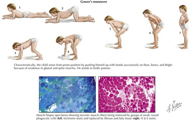
Both conditions result in marked, persistent CK elevation, generally 10 times the upper limit of normal or higher. The diagnosis can be made in more than 90% of patients by confirmation of a deletion or point mutation of the Xp21 gene locus. Analysis of dystrophin content by muscle biopsy is also a reliable diagnostic tool (see Fig. 60-1, lower).
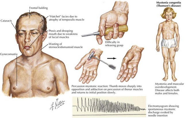

Treatment of the muscle weakness is supportive. Selected patients may benefit from surgery to retard scoliosis, respiratory support, and treatment of cardiac complications. Standard therapies with angiotensin-converting enzyme inhibitors and β-blockers are generally used for cardiac failure and arrhythmias. For drug-refractory ventricular arrhythmias, an implantable cardiac defibrillator may be beneficial. In Becker’s MD, some patients with disproportionate cardiac involvement have successfully undergone cardiac transplantation.


Emery-Dreifuss Muscular Dystrophy


Emery-Dreifuss muscular dystrophy (EDMD) is a rare, genetically heterogeneous disorder characterized by early contractures of the elbows, ankles, and posterior cervical muscles and slowly progressive muscle weakness in a scapulohumeroperoneal distribution. It is caused by mutations in nuclear proteins found in the inner muscle membrane in skeletal and cardiac muscles. Mutations in emerin cause X-linked EDMD (XL-EDMD), and mutations in lamin A/C cause autosomal-dominant EDMD (AD-EDMD). Mutation in the latter also causes limb girdle muscular dystrophy 1B. Cardiac involvement in XL-EDMD is characterized by atrioventricular (AV) conduction defects, heart block, atrial flutter, and complete atrial paralysis. The echocardiogram usually reveals a dilated right atrium; dilated or restrictive cardiomyopathy is rare. In AD-EDMD, cardiac involvement is a common complication; dilated cardiomyopathy is more common than in XL-EDMD. Supraventricular and ventricular arrhythmias, syncopal episodes, and sudden death are common in both disorders.


Clinically, patients usually have an elevated CK, but generally it is less than 10 times the normal level. However, it can be normal in patients with mutations in lamin A/C. Electromyography (EMG) shows a myopathic pattern, and muscle biopsy reveals a dystrophic picture with prominent fibrosis. Diagnosis is confirmed by genetic testing.


All patients with EDMD should have an annual ECG, echocardiogram, and Holter monitoring. Permanent pacemaker implantation is justified, either in asymptomatic patients or when the ECG becomes abnormal. In AD-EDMD, there is accumulating evidence of sudden death even in patients who have been paced. For this reason, an implantable defibrillator is more appropriate management than a pacemaker in most patients. When atrial fibrillation or atrial standstill is recognized, antithromboembolic prophylaxis with aspirin or warfarin should be considered.


Myotonic Dystrophy


Myotonic dystrophy, an autosomal-dominant MD, is a multiple-system disease. Two types of myotonic dystrophy have been identified. Type 1, or DM1, is predominantly a distal myopathy caused by an expanding trinucleotide CTG-repeat of a specific protein kinase on chromosome 19. Type 2, or DM2, is a proximal myotonic dystrophy caused by the expansion of a CCTG repeat in the zinc finger protein (ZNF9) on chromosome 3. Disease expression varies within and between affected families. Patients may have facial muscle weakness, especially of the temporalis, levator palpebrae superioris, and masseters, giving typical “cadaveric” facies. Myotonia results in delayed muscle relaxation after contraction or muscle percussion (Fig. 60-2). Systemic features may include frontal balding, cataracts, hypogonadism, insulin resistance, dysphagia, hypersomnia, pickwickian syndrome, and mental retardation. Children of affected mothers with DM1 are more likely to be severely weak and hypotonic and to have mental retardation in infancy. Progression of myotonic dystrophy is variable, with death usually resulting from aspiration pneumonia, respiratory failure, or cardiac involvement.


Cardiac manifestations are similar in both types and include conduction defects, atrial and ventricular tachyarrhythmias (occasionally causing sudden death), mitral valve prolapse, and, rarely, dilated cardiomyopathy. Anesthesia may increase the risk of AV conduction block. A yearly ECG is recommended. In high-risk families or individuals deemed to be at risk, His bundle studies and prophylactic cardiac pacemaker placement are often recommended. β-adrenergic blockers and angiotensin-converting enzyme inhibitors improve cardiomyopathic symptoms. Mexiletine, phenytoin, or other antiepileptic agents can sometimes ameliorate the myotonia. Modafinil or methylphenidate may alleviate hypersomnia.


Genetic testing can confirm the diagnosis of myotonic dystrophy. Serum CK may be elevated or normal. Myotonic discharges have a characteristic “dive bomber” sound on EMG, although this feature is not generally present in infants.


Jun 12, 2016 | Posted by in CARDIOLOGY | Comments Off on Neuromuscular Diseases and the Heart

Full access? Get Clinical Tree

Get Clinical Tree app for offline access