Natural History of Acute Venous Thrombosis


Natural History of Acute Venous Thrombosis


Sherene Shalhub and Mark H. Meissner


Acute deep vein thrombosis (DVT) and its complications, including pulmonary embolism (PE), recurrent venous thromboembolism (VTE), and the postthrombotic syndrome (PTS), are important not only as the most common preventable cause of hospital death but also as a cause of substantial long-term morbidity. The early natural history of acute DVT is characterized by a balance between factors favoring restoration of the venous lumen and recurrent thrombosis. The balance between these competing events is also related to development of PTS, the most important late complication of DVT.


Venous Thrombogenesis and the Early Natural History of Deep Vein Thrombosis


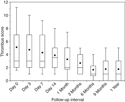
DVT is fundamentally a disease of coagulation localized to the lower extremity veins. Most of the risk factors for acute DVT are associated with some degree of hypercoagulability as a congenital, acquired, or situational risk factor (Box 1). However, most thrombi arise from multiple risk factors acting together in a synergistic fashion. Lower extremity venous thrombi originate in areas where imbalanced coagulation is localized by stasis, such as in the soleal sinuses, behind venous valve pockets, and at venous confluences. The calf veins are the most common site of origin, although 40% of proximal thrombi arise primarily in the femoral or iliac veins.


Propagation of thrombi beyond areas of stasis probably depends on the relative balance between activated coagulation and thrombolysis. Animal models have suggested that up-regulation of the selectins, with subsequent formation of microparticles derived from platelet, leukocyte, and endothelial cell membranes, may be important in amplifying the local thrombotic stimulus. Under such conditions, laminated appositional growth occurs outward from the apex as platelets are surrounded by a network of red cells, fibrin, and leukocytes. In contrast to arterial thrombi, venous thrombi are composed largely of red cells and fibrin, with relatively few platelets and leukocytes. Such early thrombi can fail to propagate, ultimately appearing as endothelialized fibrin fragments within the valve pockets.


The venous lumen is eventually reestablished following most DVT episodes. Recanalization occurs through a complex process involving intrinsic and extrinsic fibrinolysis, peripheral fragmentation, neovascularization, and retraction. A fibrinolytically active neoendothelium is regenerated soon after thrombosis. As recanalization proceeds, numerous proinflammatory factors are locally released, causing an influx of neutrophils followed by monocytes into the vein wall and thrombus. In animal models, neutrophils, which can contribute to both fibrinolysis and vein wall injury, are the first cells to appear in a thrombus. A predominantly monocyte infiltrate follows and peaks on day 8 after thrombosis. The monocyte plays a central role in late thrombus organization and recanalization, serving as a source of both fibrinolytic and cytokine mediators. Leukocytes are simultaneously responsible for a number of changes occurring in the vein wall. An inflammatory process involving profibrotic growth factors, collagen deposition, and matrix metalloproteinase (MMP) expression and activation can mediate late wall fibrosis and reduced vein wall compliance.


Recanalization has been demonstrated clinically in several series employing serial duplex ultrasonography. Most recanalization occurs within the first 6 to 12 weeks after the acute event, and the original thrombus load is reduced by 50% to 60% within 6 to 9 months of thrombosis (Figure 1). The degree of activated coagulation and fibrinolytic inhibition are independent predictors of recanalization. Clinically, greater recanalization has been associated with thrombosis provoked by a transient risk factor, and malignancy and the presence of a permanent risk factor have been associated with less complete recanalization. In comparison to treatment with warfarin, greater recanalization has also been reported following 3 months of treatment with low-molecular-weight heparin.


Stay updated, free articles. Join our Telegram channel

Aug 25, 2016 | Posted by in CARDIOLOGY | Comments Off on Natural History of Acute Venous Thrombosis

Full access? Get Clinical Tree

Get Clinical Tree app for offline access