Narrowed Aorta
Gregory Kicska, MD, PhD
DIFFERENTIAL DIAGNOSIS
Common
Coarctation of Aorta
Pseudo-coarctation
Less Common
Large Vessel Vasculitis
Rare but Important
Extrinsic Aortic Compression
ESSENTIAL INFORMATION
Key Differential Diagnosis Issues
Focal outer diameter narrowed in coarctation or vasculitis
Diffuse outer diameter narrowing can occur in vasculitis
Normative data should be consulted to exclude common pitfall of misinterpreting normal aortic dimension for small body as abnormal
For > 45 years, aorta considered abnormally small if diameter at level of main pulmonary artery < 24 mm for ascending and < 18 mm for descending aorta
Larger diameters may still be abnormal if older age, larger body surface area (BSA), or male
Helpful Clues for Common Diagnoses
Coarctation of Aorta
Focal narrowing occurs below ductus arterious in adults, at ductus arterious with arch hypoplasia in neonates
Dilated collaterals (intercostal, internal thoracic) indicate hemodynamically significant coarctation
Treated coarctation patients can have restenosis
Associated with bicuspid aortic valve, Turner syndrome, Marfan syndrome
Pseudo-coarctation
Redundant aorta with narrowing distal to left subclavian origin without hemodynamic effect
No rib notching, cardiomegaly, or collateral vessels
Dilated brachiocephalic artery and high arch often present
Helpful Clues for Less Common Diagnoses
Large Vessel Vasculitis
Variable segment length narrowing
Branch vessel narrowing is common
Periaortic thickening and enhancement
Periaortic FDG uptake implies active disease
< 40 years implies Takayasu, > 40 years implies giant cell arteritis
Helpful Clues for Rare Diagnoses
Extrinsic Aortic Compression
Retroperitoneal fibrosis, neurofibromatosis, sarcomas, other neoplasms
Image Gallery
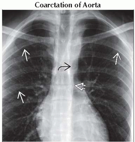 PA radiograph shows subtle areas of rib notching
 . Note the rapid narrowing of the proximal descending thoracic aorta . Note the rapid narrowing of the proximal descending thoracic aorta  , which then returns to normal size more distally , which then returns to normal size more distally  . .Stay updated, free articles. Join our Telegram channel
Full access? Get Clinical Tree
 Get Clinical Tree app for offline access

|