Myocarditis

22 Myocarditis


Myocarditis is an inflammatory process that can involve one or more components of the myocardium including cardiomyocytes, the interstitium, and the coronary vasculature. This inflammatory process may result from infectious processes, responses to pharmacologic or toxic agents, hypersensitivity reactions, or physical damage. Myocarditis may also be a cardiac manifestation of a systemic disease.


The clinical course of myocarditis is as diverse as its etiologies. Most patients have a subclinical, self-limited course, but myocarditis may also have fulminant, acute, or chronic presentations. The burden of myocarditis as a clinical entity is difficult to ascertain, at least in part because of its diversity and the elusiveness of diagnosis; for similar reasons, the ideal diagnostic and therapeutic approach to myocarditis has been elusive. The future is likely to be more promising. Recent data have established a causal link between the chronic effects of viral myocarditis and dilated cardiomyopathy. New treatments for dilated cardiomyopathy and heart failure have focused on immunomodulating therapy partly based on this knowledge. Further elucidation of the pathogenesis of myocarditis will probably affect the management of left ventricular (LV) dysfunction and heart failure.


Etiology and Pathogenesis


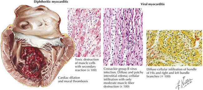
In North America and Europe, the majority of cases of myocarditis probably result from viral infection. Many viruses have been associated with myocarditis (Box 22-1). Initial serologic studies suggested that enteroviruses, such as coxsackie B, are common causes of viral myocarditis. However, the application of direct molecular techniques to endomyocardial biopsy specimens, and perhaps changing epidemiology, has led to increasing recognition of adenoviruses, parvovirus, and hepatitis C as etiologic agents. In HIV infection, there is often evidence of myocarditis when cardiac decompensation occurs, although it is unclear whether HIV or opportunistic infections are responsible.


The molecular mechanisms of myocardial injury in viral myocarditis remain incompletely understood. The initial phases of injury probably depend on viral attachment to myocytes and direct cell damage by the virus, resulting in myocyte necrosis. The finding of a common membrane receptor for adenoviruses and coxsackieviruses supports this hypothesis and the preponderance of these viruses as causative agents. Following the initial injury, host immune response to the virus probably has an important role in myocardial injury. Animal models have shown that after the initial phase of entry and proliferation of the virus in the myocyte cytoplasm, inflammatory cells (including natural killer cells and macrophages) infiltrate with subsequent release of proinflammatory cytokines. T lymphocytes are activated through classic cell-mediated immunity. Cytotoxic T cells recognize viral protein fragments on the cell surface in a major histocompatibility complex-restricted manner. Molecular mimicry can occur when antigens intrinsic to the myocyte cross-react with viral peptides, inducing persistent T-cell activation. Cytokines, including tumor necrosis factor, interleukin (IL)-1, IL-2, and interferon γ have been identified as important mediators of chronic inflammatory disease. These cytokines can cause myocyte damage, resulting in fewer contractile units with a resulting worsening of systolic function. Autoantibodies to myocyte components are often found in patients with myocarditis, although most studies measuring autoantibody levels were in patients with idiopathic dilated cardiomyopathy. Even so, it is likely that cellular immunity has more of a role in the pathogenesis of myocarditis than does humoral immunity.


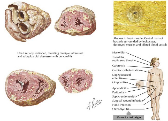
Rarely, bacterial infections, through spread from endogenous sources (Fig. 22-1), can produce focal or diffuse myopericarditis. One of the earliest recognized causes of myocarditis was diphtheria. Up to 20% of diphtheria patients have cardiac involvement, and myocarditis is the leading cause of death with this infection. The toxin produced by the diphtheria bacillus injures myocardial cells (Fig. 22-2). In South and Central America, the most common cause of infectious myocarditis is the protozoan Trypanosoma cruzi—the causative agent of Chagas’ disease.


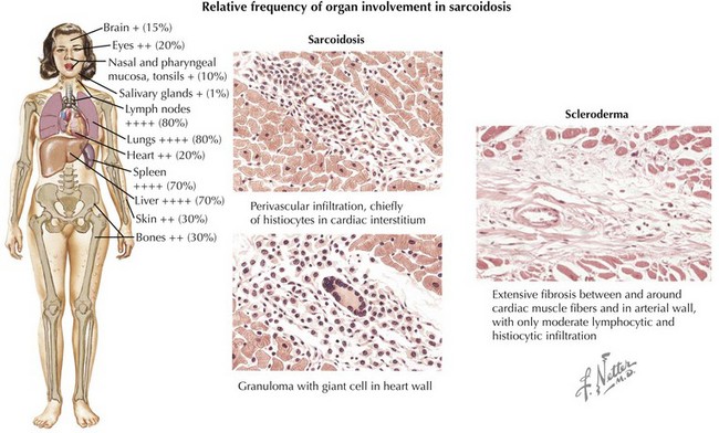
Sarcoidosis, a systemic granulomatous disorder of unknown etiology, involves the myocardium in at least 20% of cases. Cardiac involvement ranges from a few scattered lesions to extensive involvement (Fig. 22-3). As a result, endomyocardial biopsy may be diagnostic but is frequently unreliable in confirming myocarditis. Giant cell myocarditis is a rare but highly lethal form of myocarditis of suspected immune or autoimmune etiology that may be associated with other inflammatory conditions such as Crohn’s disease. Although the cumulative studies on immunosuppressive therapy for myocarditis are not positive (see below), the above causes of myocarditis do often respond to immunosuppression. Peripartum cardiomyopathy has been associated with a greater than 50% rate of myocarditis on endomyocardial biopsy, although the etiology remains unknown.


Hypersensitivity reactions resulting in myocarditis are characterized by eosinophilia and a perivascular infiltration of the myocardium by eosinophils and leukocytes. Any drug may cause hypersensitivity myocarditis, but clinically this condition is rarely recognized. Therefore, a high index of suspicion should be maintained.

Stay updated, free articles. Join our Telegram channel

Jun 12, 2016 | Posted by in CARDIOLOGY | Comments Off on Myocarditis

Full access? Get Clinical Tree

Get Clinical Tree app for offline access