Myocardial Protection for Neonates and Infants


Myocardial Protection for Neonates and Infants


Paul J. Chai

Barry D. Kussman


INTRODUCTION

The goal of myocardial protection in pediatric cardiac surgery is to allow for a technically perfect repair without cardiac injury. Essentially all intracardiac repairs in neonates and infants require a period of ischemia to provide an immobile and bloodless field (1). Whatever technique is used, the principle is to maintain a favorable balance between myocardial oxygen supply and demand. It is important to be aware that myocardial protection is not limited to the period of ischemia during cardiopulmonary bypass (CPB), and includes optimum management of the patient preoperatively, intraoperatively before and after any myocardial ischemia, and in the postoperative period. Multiple factors in addition to myocardial preservation influence postoperative cardiac output and outcome.

Pediatric myocardial protection differs from that of adults in a number of significant ways. The immature heart has differences in structure and function, and as the myocardium matures, fundamental changes occur that directly influence the ability of the heart to withstand periods of ischemia and injury. Although coronary artery occlusion is rare, many congenital cardiac anomalies are associated with impaired myocardial function (“stressed” myocardium) before reparative surgery. Myocardial protection techniques must be tailored to the age of the patient, the cardiac physiology, and the complexity of the surgical procedure in order to achieve the best outcomes.


BIOLOGY OF MYOCARDIAL INJURY AND PROTECTION

Factors to consider with respect to the biology of myocardial injury and protection are listed in Table 29.1.


Physiologic Differences between Immature and Mature Myocardium

The precise age of crossover from the immature myocardium of the neonate and infant to the mature myocardium of the child and adult is not well defined (1). Differences in structure and function between immature and mature hearts have implications for myocardial protection during ischemia. The normal neonatal myocardium is generally considered to be more resistant to ischemia than mature myocardium (2,3,4,5). However, the “stressed” myocardium is thought to be more sensitive to ischemia. The physiologic differences allowing immature myocardium greater tolerance to ischemia have been reviewed by Doenst et al. and are considered below (6) (Table 29.2).


TABLE 29.1. Biology of myocardial protection


Immature vs. mature myocardium


Energy substrate


Calcium metabolism


Enzyme activity


Catecholamine sensitivity


Ischemic preconditioning


Normal vs. stressed heart


Hypoxia and cyanosis


Heart failure


Ventricular hypertrophy (pressure and volume overload)


Ischemia-reperfusion injury


Coronary vascular dysfunction


Nonischemic causes of myocardial injury


Ventricular distension


Retraction injury


Myocardial edema


Ventriculotomy


Coronary artery injury


Energy Substrate

Myocardium derives its energy production, adenosine triphosphate (ATP), from various sources that include free fatty acids, glucose, glycogen, lactate, pyruvate, ketones, and amino acids. In the adult heart under aerobic conditions, 95% of energy production is from oxidation of long-chain fatty acids (7). In contrast, glucose is the main substrate for the neonatal heart, and is supplemented by oxidation of fatty acids, lactate, ketones, and amino acids (8). The timing of the shift in substrate preference from glucose to fatty acids varies
between species and is thought to be due to upregulation of 5′-adenosine monophosphate-activated protein kinase (9). During this period of development, the neonatal myocardium shows a progressive decline in glucose uptake which can be stimulated by insulin, that is, insulin resistance and a much greater capacity to store glycogen (10). The greater ability of the immature myocardium to utilize anaerobic glycolysis may partially account for the greater tolerance to ischemia. Laboratory and clinical studies in adults have found that enhanced glucose uptake and oxidation is associated with enhanced functional myocardial recovery, despite normalization of fatty acid oxidation (6).


TABLE 29.2. Physiologic differences between pediatric and adult myocardium and the potential impact of these differences on ischemia tolerance of the pediatric heart


Pediatric


Adult


Potential impact on ischemia tolerance in the pediatric heart


Preferred substrate for adenosine triphosphate production


Glucose


Fatty acids


Increase


Glycogen content


High


Low


Increase


Insulin sensitivity


Impaired


Normal


a


Calcium handling (intracellular)


Impaired


Normal


a


Calcium sensitivity


Increased


Normal


Decreasea


Antioxidant defense


Low


High


Decrease


5′ nucleotidase


Low


High


Increase


Catecholamine sensitivity


Low


Normal


a


Ischemic preconditioning


Absent


Present


a


a Potential effect unknown.


Reprinted from Doenst T, Schlensak C, Beyersdorf F. Cardioplegia in pediatric cardiac surgery: do we believe in magic? Ann Thorac Surg 2003;75: 1668-77, with permission.


Calcium Metabolism

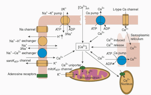
Immature myocardium is significantly more sensitive to and dependent on extracellular calcium than adult myocardium (Fig. 29.1). The sarcoplasmic reticulum in immature myocardium is less well developed and the sarcoplasmic reticular Ca2+-ATPase activity is also reduced. The result is a reduced storage capacity for calcium, reduced calcium uptake into the sarcoplasmic reticulum, and decreased calcium release following ryanodine receptor activation. Therefore, there is greater dependence on movement of calcium from the extracellular to the intracellular space (11,12,13,14). The decreased capacity for calcium sequestration may explain why the immature myocardium is susceptible to postischemic calcium overload. Several studies have found adverse effects with cardioplegia solutions containing normal or high concentrations of calcium (15,16). Most cardioplegia solutions in use currently contain very low levels of calcium (17,18,19,20,21).


Enzyme Activity

Two enzyme systems seem to be important for myocardial protection during periods of ischemia: the antioxidant system and 5′ nucleotidase. The antioxidant system includes superoxide dismutase, catalase, and glutathione reductase and is responsible for scavenging the oxygen-derived free radicals generated during reperfusion. Reactive oxygen species (ROS), which comprise superoxide, hydrogen peroxide, hydroxyl radical, and lipid peroxides, cause peroxidation of phospholipids in cell membranes leading to loss of cellular integrity and function. In addition to enzyme depletion during long periods of ischemia, the activity of this enzyme system is reduced in immature myocardium (22). Another study, however, found significantly increased baseline catalase activity and reduced xanthine oxidase activity in newborn rat heart (23). Neonates with tetralogy of Fallot have a significant reduction in the activity of antioxidant enzymes (24), and may be at even greater risk.

The enzyme 5nucleotidase catalyzes the conversion of adenosine monophosphate (AMP) to adenosine. While AMP is unable to pass through the cell membrane, adenosine passes easily and is rapidly lost to the extracellular space, thereby depleting the adenine nucleotide pool (AMP, ADP, and ATP). The size of this adenine nucleotide pool is important for, though not predictive of, postischemic recovery of the myocardium (25,26,27). The 5nucleotidase system is reduced in immature myocardium and may be an additional explanation as to why immature myocardium is more tolerant to ischemia (28). If the pool is depleted more than 50%, immediate full recovery of contractile function is not possible (27,29,30).


FIGURE 29.1. Sources of calcium regulation. Calcium entry via the L-type Ca2+ channel causes Ca2+ release from the sarcoplasmic reticulum (SR) via the ryanodine receptor (Ca-induced Ca release) and activation of contraction. Calcium is pumped back into the SR by SR Ca2+-ATPase and extruded from the cell by activating the Na+-Ca2+-exchanger to allow relaxation. Calcium can also enter mitochondria via a Ca2+ uniporter. (Reprinted from Levitsky S, McCully JD. Myocardial protection. In: Sellke FW, del Nido PJ, Swanson SJ, eds. Sabiston & Spencer surgery of the chest. 8th ed. Philadelphia, PA: Saunders Elsevier, 2010:977-998, Figure 63-4 (p. 980), with permission.)


Catecholamine Sensitivity

The sensitivity to catecholamines is decreased in immature hearts. An in vitro study found evidence to suggest that this is due to functionally incomplete coupling of myocardial β-adrenergic receptors to adenylate cyclase at birth (31). In contrast, the kinetics of cyclic AMP hydrolysis and the inhibitory potential of phosphodiesterase inhibitors were not affected by age. Whether this decreased catecholamine sensitivity plays a role in tolerance to ischemia is uncertain.


Ischemic Preconditioning

Ischemic preconditioning can be defined as the adaptive mechanism induced by a brief period of reversible ischemia increasing the heart’s resistance to a subsequent longer period of ischemia (32). Although protective for older age groups, ischemic preconditioning was not found to be effective in the newborn rat heart (33). Conversely, chronic hypoxia is associated with reduced tolerance to myocardial ischemia.


Normal versus Stressed Myocardium

Although normal neonatal myocardium may be more tolerant to an ischemic insult than adult myocardium, this tolerance does not seem to be present in hearts that have been stressed by exposure to severe hypoxemia, chronic cyanosis, ventricular hypertrophy, or refractory heart failure (15,34,35,36,37,38,39). These myocardial stressors are frequently present in neonates and infants and are associated with depletion of high-energy phosphates, glycogen, and Kreb’s cycle intermediates (40). Volume and pressure loading increase myocardial oxygen demand, and ventricular hypertrophy can result in relative hypoperfusion of the subendocardium. Cyanotic lesions with decreased pulmonary blood flow tend to have aortopulmonary collaterals that during CPB increase left heart return, rewarm the heart, and washout cardioplegia. Stressed myocardium translates into greater depression of systolic and diastolic function following ischemia and reperfusion injury. As myocardial energy stores in normal piglet hearts have been found to differ between individuals, some unstressed hearts may be more susceptible to ischemia (41).


Ischemia-Reperfusion Injury

The heart is an obligate aerobic organ with injury to the myocardium occurring during both ischemia and reperfusion. Oxygen and the production of ATP are necessary for the external mechanical work of contraction and basal metabolism (unloaded contraction) (42). Myosin ATPase is required for the development of wall tension, sarcoplasmic Ca2+-ATPase for the sequestration of calcium, and Na+-K+-ATPase for the maintenance of the membrane potential. Experimentally, myocardial oxygen consumption (MVo2) in different states in the neonate differs somewhat from what studies in adult hearts have shown. MVo2 (expressed in mL of O2 per 100 g of ventricular
tissue per minute) averaged 6.7 mL in the working state (adult 8 mL), 3.2 mL in the empty beating state (adult 5.6 mL), 1.3 mL in the potassium-arrested heart at 37°C (adult 1.1 mL), 0.37 mL in the hypothermic (15°C) heart, and 0.32 mL in the hypothermic (15°C) potassium-arrested heart (43,44).

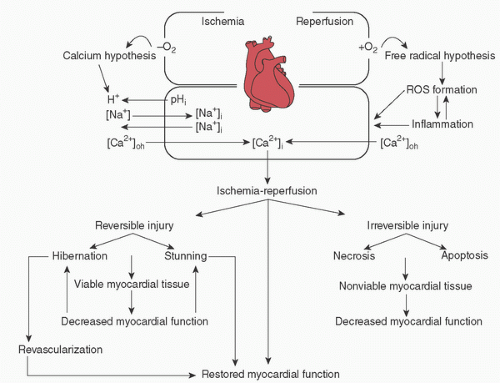
Ischemia-reperfusion injury involves multiple cellular and extracellular processes and is reviewed in detail elsewhere (18). Briefly, ischemia and reperfusion are associated with the depletion of ATP, intracellular acidosis, intracellular calcium overload, inflammation, myocardial edema, generation of ROS, and endothelial dysfunction. Reversible ischemia-reperfusion injury may occur as “stunning” (contractile dysfunction persisting after normal or near-normal reperfusion in the absence of cell damage) or “hibernation” (reversible chronically reduced contractile function).

Putative mechanisms involved in ischemia-reperfusion injury are shown in Figure 29.2. The calcium hypothesis is based on the inability of the myocyte to regulate intracellular and intraorganellular calcium concentration such that increased calcium activates a cascade of events resulting in cell dysfunction, cell injury, and/or cell death. The free-radical hypothesis suggests that accumulation of ROS during the early stages of reperfusion causes myocyte injury through peroxidation of the cellular phospholipid layers. The lethal reperfusion injury hypothesis has been described in adults in the setting of acute coronary occlusion and refers to the death of myocardial cells that were viable immediately before reperfusion (45). It presupposes that during reperfusion the biochemical and metabolic changes compound the changes produced during the period of ischemia and interact with each other to mediate cardiomyocyte death through the opening of the mitochondrial permeability transition pore (PTP) and the induction of cardiomyocyte hypercontracture (46). The mitochondrial PTP is a nonselective channel of the inner mitochondrial membrane whereby channel opening minimizes the mitochondrial membrane potential leading to uncoupling of oxidative phosphorylation, ATP depletion, and cell death (47).


FIGURE 29.2. Mechanisms of ischemia-reperfusion injury. Putative mechanisms of the calcium and free-radical hypotheses and inflammation in the generation of ischemia-reperfusion injury. ROS, reactive oxygen species. (Reprinted from Levitsky S, McCully JD. Myocardial protection. In: Sellke FW, del Nido PJ, Swanson SJ, eds. Sabiston & Spencer surgery of the chest. 8th ed. Philadelphia, PA: Saunders Elsevier, 2010:977-998, Figure 63-3 (p. 980), with permission.)


Coronary Vascular Dysfunction

Endothelial cells regulate microcirculatory vascular tone by promoting vascular relaxation and inhibiting platelet function (48). Ischemia-reperfusion and the systemic inflammatory response to CPB are associated with endothelial injury and impaired release of nitric oxide (NO), prostacyclin, and adenosine. NO is a potent smooth muscle relaxant and inhibitor of platelet and neutrophil adhesion, and is thought to be an important component in the recovery of myocardial
(and pulmonary) function as a result of ischemia-reperfusion injury and the associated endothelial dysfunction (49). In neonatal lambs, the addition of L-arginine (NO precursor) or nitroglycerine (NO donor) during reperfusion resulted in significantly higher preload recruitable stroke work and cardiac index (50). A clinical practice at Boston Children’s Hospital is to start a nitroglycerine infusion (1 µg/kg/min) 5 to 10 minutes before removal of the aortic cross-clamp and continue it until the end of rewarming.


Nonischemic Causes of Myocardial Injury


Ventricular Distension

There is a general feeling among pediatric cardiac surgeons that immature myocardium is significantly more vulnerable to stretch injury. Stretch injury typically occurs with overdistension of the left heart and excessive retraction on myocardial tissues, causing ventricular injury and contractile dysfunction. It can also lead to distension of the left atrium and pulmonary veins, resulting in elevated pulmonary transcapillary pressure and development of significant pulmonary edema and pulmonary dysfunction. Ventricular distension can occur with aortic valve insufficiency prior to cross-clamping the aorta. Excessive pulmonary venous return in the setting of reduced heart rate and myocardial function, ventricular fibrillation during cooling, or multiple aortopulmonary collaterals can also cause distension of the left ventricle.

The cardiac surgeon needs to be acutely aware of instances when distension of the left heart can occur. Slow cooling with maintenance of ventricular ejection until application of the aortic cross-clamp is important in the setting of aortic insufficiency. Inspection and palpation of the main pulmonary artery and left ventricle, as well as massage of the left ventricle, are important. Left heart venting should be performed either through the right superior pulmonary vein or through an atrial septal defect to keep left atrial and ventricular pressures low. The safest time to insert a left atrial vent is after the aortic cross-clamp is applied in order to prevent entrainment of air into the left ventricle and ejection into the systemic circulation. Depending on the etiology, pulmonary venous return can be reduced by preoperative coil occlusion of collaterals, shunt, or PDA ligation after commencement of CPB, or opening of the pulmonary artery on CPB. An instant measure to reduce left ventricular distension is having the perfusionist quickly drop the flow rate and perfusion pressure until the issue is resolved. Removal of the left heart vent can also result in entrainment of air, and so should only occur when the heart is contracting and the patient is on partial bypass with the left heart filled with blood above atmospheric pressure.


Retraction Injury

Excessive retraction to improve exposure can injure the myocardium and conduction system. Retraction force of assistants needs to be monitored and adjusted as necessary.


Myocardial Edema

Myocardial edema can result from ischemia and reperfusion injury, delivery pressure of cardioplegia, cardioplegia osmolarity and chemical composition, excessive hemodilution, the inflammatory response to CPB, impaired myocardial lymphatic drainage, and handling of the myocardium (18,51). Myocardial edema causes impaired diastolic function.


Ventriculotomy

A ventriculotomy is a component of some surgical repairs and may cause direct myocardial injury and dysfunction. Typical operations include repair of tetralogy of Fallot, some ventricular septal defects, and placement of a right ventricle-to-pulmonary artery conduit.


Coronary Artery Injury

Injury to the coronary arteries may occur during reimplantation (arterial switch operation, aortic root/ascending aorta surgery), incisions in the anterior wall of the right ventricle (some cases of tetralogy of Fallot), or mitral valve surgery. Reoperations also pose a risk of coronary artery injury.


TECHNIQUES FOR MYOCARDIAL PROTECTION

There is no clear consensus concerning the optimal strategy for myocardial protection in the neonate and infant. Although strategies and composition of cardioplegia vary significantly from center to center, hypothermia for reduction of metabolic activity and cardioplegia for electrical and contractile arrest is the mainstay. Published studies often demonstrate conflicting results, which make it difficult to recommend a “best” method for myocardial protection.


Preischemic Management

Patients who are well resuscitated before surgically-induced ischemia generally have a better outcome (1). This is particularly relevant for the neonate or infant with a “stressed” heart. The goal is achievement of a normal metabolic state with reversal of tissue ischemia as manifested by hemodynamic stability, normal renal and hepatic function, and appropriate blood pH and lactate levels. Apart from newborns with obstructed total anomalous pulmonary venous connection, this can be accomplished by administration of a prostaglandin E1 infusion (alprostadil 0.01-0.05 µg/kg/min) to maintain or reestablish ductal patency, endotracheal intubation and mechanical ventilation, fluid administration, and inotropic support.

Maintenance of blood pressure and myocardial blood flow in the operating room is also important. When the pulmonary and systemic circulations are connected, lowering of the pulmonary vascular resistance by general anesthesia and mechanical ventilation leads to diastolic runoff, systemic hypotension, and decreased myocardial blood flow (1

Only gold members can continue reading. Log In or Register to continue

Stay updated, free articles. Join our Telegram channel

Jun 7, 2016 | Posted by in RESPIRATORY | Comments Off on Myocardial Protection for Neonates and Infants

Full access? Get Clinical Tree

Get Clinical Tree app for offline access