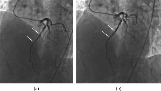
Percutaneous coronary angiography showed myocardial bridging in LAD with systolic narrowing of lumen diameter with 50% of compression in systole and normal in diastole (Figure 35.2). The remaining coronary arteries were normal.
Figure 35.2 Percutaneous coronary angiography showed myocardial bridging (arrows) in left anterior descending during left ventricle systole (a) and diastole (b).
Discussion
Stay updated, free articles. Join our Telegram channel

Full access? Get Clinical Tree


