Mucoepidermoid Carcinoma
Presentation
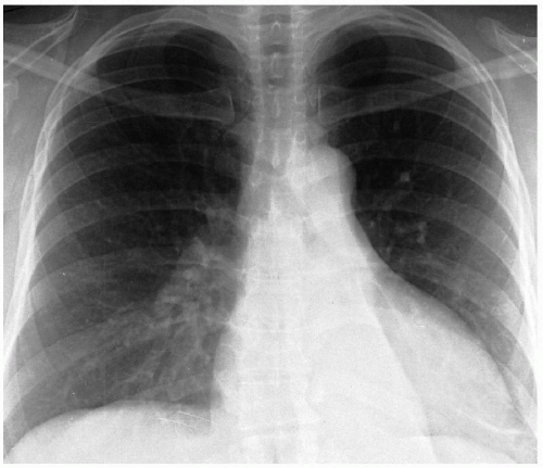
A 40-year-old man with no significant past medical history is referred by a primary medical physician for evaluation of an abnormal chest x-ray finding. The patient reports an episode of chest pain accompanied by fever and mild dyspnea 3 years ago. Antibiotics and analgesics were prescribed at that time, which resulted in significant improvement. At this time, the patient experiences wheezing and a dry cough upon lying flat. He was evaluated by a pulmonologist who recommended inhalers. With inhaler therapy, the symptoms did not improve, and the cough became productive of thick white sputum, occasionally blood tinged.
Recommendation
Computed tomography (CT) scans of the chest.
▪ CT Scans
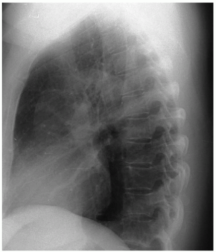
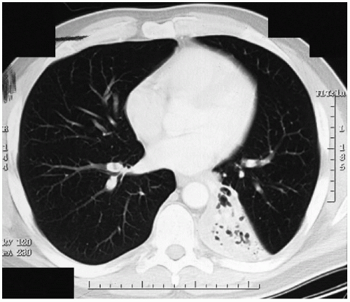 Figure 54-3 |
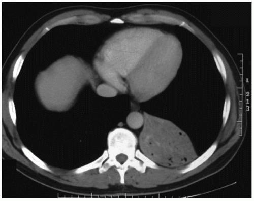 Figure 54-4 |


