Mucinous Adenocarcinoma


Mucinous Adenocarcinoma


Allen P. Burke, M.D.

Adina Paulk, M.D.


Introduction and Terminology

The classification of mucinous adenocarcinomas of the lung is complicated and has been described as “difficult and somewhat arbitrary”1 as well as “controversial.”2 The difficulties stem from the fact that many mucinous carcinomas show mixed patterns (including so-called enteric; see Chapter 77), and there is a variable in situ or lepidic component, a feature that in lung cancers has long caused confusion. Furthermore, mucin takes many forms, including intracellular (in goblet, columnar, and signet ring cells) as well as extracellular, often entrapping small tumor nests (a feature characterizing “colloid” carcinoma).

A general concept maintains that there are two cell types of origin for lung adenocarcinomas: pneumocytes with either type II pneumocyte or Clara cell morphology3 or a mucin-producing cell corresponding to bronchial goblet cells (see chapter 75).4 It is generally recognized as well that mucinous adenocarcinomas are TTF-1 negative, although there are many mixed tumors with a subpopulation of TTF-1 positive non-mucin-secreting cells.

For purposes of this chapter, only adenocarcinomas with extensive extracellular mucin are considered. Solid adenocarcinomas with prominent intracellular mucin vacuoles or signet ring morphology are considered as solid (nonmucinous) adenocarcinomas and for the most part are TTF-1 positive.4,5

In contrast to nonmucinous carcinomas, mucinous adenocarcinomas of the lung are not often entirely in situ. Because few data exist for large noninvasive tumors, any mucinous adenocarcinoma >3 cm with an in situ (lepidic) growth pattern is considered invasive by the World Health Organization classification6 (Table 76.1).


Mucinous Adenocarcinoma In Situ and Minimally Invasive Adenocarcinoma


Microscopic Findings

Unlike nonmucinous adenocarcinomas, the lepidic spread of mucinous adenocarcinoma in situ is characterized by completely normal alveolar septa without any fibrotic thickening (Fig. 76.1). The tumor cells are more often columnar than goblet, follow the contours of the alveolar septa, and show uniform polarity, apical intracellular mucin (as nuclei are parallel and basally located), and a fine brush border (Fig. 76.2). Alternatively (and in mixed mucinous and nonmucinous tumors), cells may be more cuboidal, with variable numbers of goblet cells (Figs. 76.3 and 76.4). The alveolar spaces are typically filled with mucin and scattered inflammatory cells. By definition, there are no detached intra-alveolar tumor cell clusters in pure adenocarcinoma in situ or minimally invasive adenocarcinoma, and the lesion is single. If there is an invasive component, it is by definition <5 mm (minimally invasive adenocarcinoma) and consists entirely of acinar, solid, or papillary growth without necrosis or lymphatic invasion.


Immunohistochemical Findings

Mucinous adenocarcinoma in situ is typically TTF-1 negative5 although older reports using the term “bronchioloalveolar carcinoma” have shown up to a 30% positivity rate.1,4 It is often difficult to distinguish tumor cell nuclei from native pneumocytes lining the alveolar spaces, especially if the tumor nuclei are compressed toward the cell base (goblet cell morphology). Other pneumocyte-related antigens
such as napsin-A and surfactant proteins (such as SP1) are negative as well.1 Expression of MUC5A5 (secreted tracheobronchial mucin) is present, and MUC2 (secreted intestinal mucin) is absent.4 Mucinous adenocarcinoma in situ is negative for CDX2.1


FIGURE 76.1 ▲ Mucinous adenocarcinoma in situ. The alveolar septa are typically thin and normal in appearance.


Invasive Mucinous Adenocarcinoma


Terminology

The term “mucinous adenocarcinoma” or “invasive mucinous adenocarcinoma” is currently considered a “variant” of adenocarcinoma by the International Association for the Study of Lung Cancer, American Thoracic Society, and European Respiratory Society (and hence the World Health Organization).6 Because the term replaced the previous “mucinous bronchioloalveolar carcinoma,” it presumably denotes the presence of a prominent in situ component. However, prior to the abolition of “bronchioloalveolar carcinoma,” the term was usually used interchangeably with “colloid carcinoma.”1 Mucinous adenocarcinomas without a predominant lepidic growth and that do not fit the criteria for colloid carcinoma do exist; however, these do not have a comfortable niche in the current WHO schema, and will also be considered here.


FIGURE 76.2 ▲ Mucinous adenocarcinoma in situ, columnar cells. The lining cells are often columnar, with pseudostratified parallel nuclei at the base. A. Routine stain showing apical mucin vacuoles and intra-alveolar mucin. B. A periodic acid-Schiff stain demonstrates diffuse cytoplasmic mucin, with apparent basal sparing because of the nuclei.


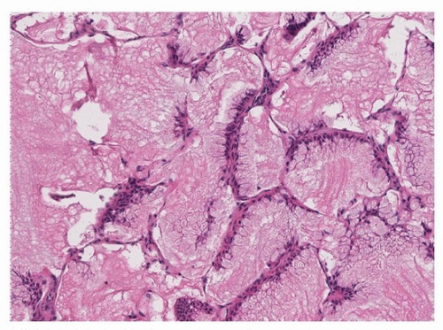
FIGURE 76.3 ▲ Mucinous adenocarcinoma in situ, goblet cell predominance. The lepidic component may have a predominance of goblet cells, with flattened basal nuclei. The tumor cell apices seem to be extruding mucin into the alveolar spaces.

For historical reasons, the “patterns” of carcinoma growth (solid, acinar, papillary, and micropapillary) have not been applied to the invasive areas of mucinous adenocarcinomas, along with adenocarcinomas of other “variant” histology.6,8 However, each of these patterns occurs in mucinous adenocarcinomas, as well as in mixed mucinous and nonmucinous adenocarcinomas. To be considered mucinous, it is generally accepted that the predominant component (>50%) of the tumor be composed of goblet or columnar cells with intracytoplasmic mucin; tumors with lesser degrees of mucinous differentiation are included under conventional adenocarcinoma and typed by growth pattern.6,8

Only gold members can continue reading. Log In or Register to continue

Stay updated, free articles. Join our Telegram channel

Aug 19, 2016 | Posted by in CARDIOLOGY | Comments Off on Mucinous Adenocarcinoma

Full access? Get Clinical Tree

Get Clinical Tree app for offline access