Mitral Valve Disease

36 Mitral Valve Disease


Mitral valve leaflets consist of thin, pliable, fibrous material. The two leaflets—anterior and posterior—open by unfolding against the ventricular wall and close by apposition when the pressure in the left ventricle becomes greater than that in the left atrium. Mitral stenosis occurs when the mitral valve leaflets become stiffened, calcified, and unable to open completely during diastole. This process often involves the chordae tendineae in addition to the mitral valve leaflets. Mitral valve regurgitation occurs when the leaflets are unable to close completely in systole. In the United States, more than 20,000 patients annually require surgery for manifestations of mitral stenosis and mitral regurgitation, and thousands more require monitoring and treatment.


Etiology and Pathogenesis
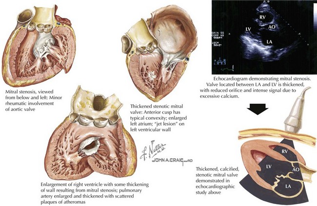


Mitral Stenosis


Rheumatic fever is responsible for a majority of cases of mitral stenosis. The initial infection and its sequelae result in thickened valve leaflets and fusion of the commissure between the leaflets. Chordae tendineae are also affected and become thickened, fused, and shortened. Most valves that are affected by rheumatic fever show abnormalities of all these structures. Few patients with rheumatic mitral valve disease have pure mitral stenosis; most have a combination of stenosis and regurgitation, and many have aortic and tricuspid involvement. Approximately two thirds of mitral stenosis cases in the United States occur in women.


The normal mitral valve cross-sectional area in diastole is 4 to 6 cm2. Blood flow is impaired when the valve orifice is narrowed to less than 2 cm2, creating a pressure gradient with exertion. A valve area smaller than 1 cm2 is considered critical mitral stenosis and results in a significant pressure gradient across the valve at rest with chronically increased left atrial (LA) pressures (Fig. 36-1).


Chronically increased LA pressures associated with mitral stenosis, along with ongoing rheumatic inflammation, result in LA enlargement and a predisposition for atrial fibrillation. Valves affected by mitral stenosis are also vulnerable to recurrent thrombosis and implantation of bacteria that lead to infective endocarditis.


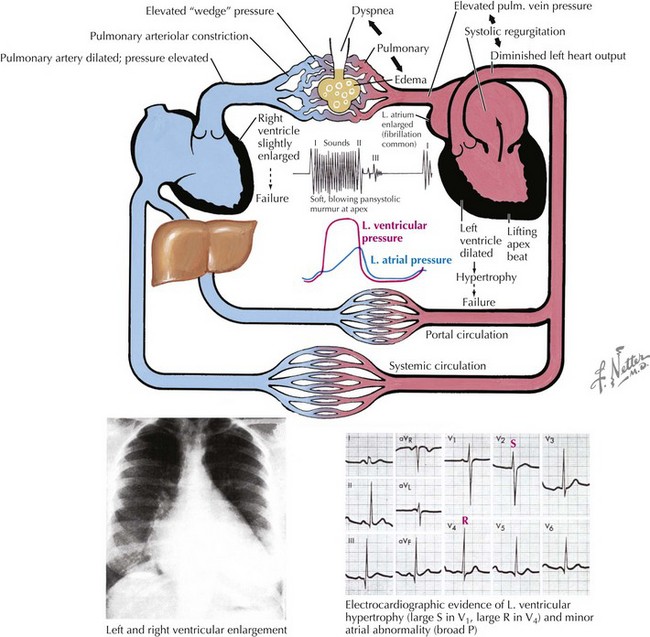
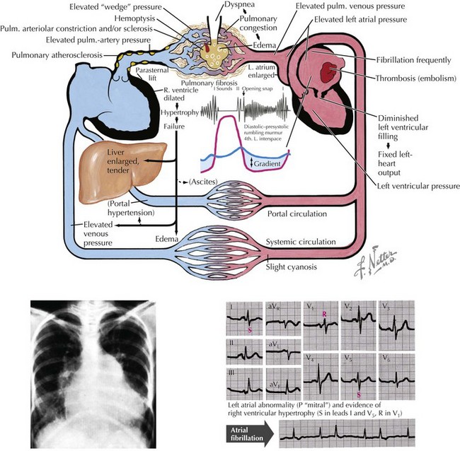
The hemodynamic effects of chronic mitral stenosis include pulmonary venous and arterial hypertension; right ventricular (RV) hypertrophy, dilation, and failure; peripheral edema; tricuspid regurgitation; ascites; and hepatic injury with cirrhosis (Fig. 36-2).


Mitral Regurgitation


Numerous etiologies contribute to mitral regurgitation, including mitral valve prolapse, rheumatic heart disease, bacterial or fungal endocarditis, and certain collagen-vascular diseases. Dysfunction of any component of the mitral apparatus can cause mitral regurgitation. Mitral regurgitation also frequently occurs in the absence of primary mitral valve disease in patients with cardiomyopathy and ventricular dilation. When the cause of mitral regurgitation is primarily a valve defect, valve repair or replacement can correct the mitral regurgitation and improve long-term prognosis. When the valve leaks because the ventricle is dysfunctional and dilated, mitral repair or replacement may have little or no effect on symptoms or prognosis.


With mitral regurgitation, blood is discharged during systole into the left atrium in addition to traveling its usual route through the aortic valve and into the aorta. If the regurgitant volume is large, LA remodeling occurs with dilation to accommodate increased volumes without intolerable LA hypertension (Fig. 36-3). Over time, as an increasing fraction of ventricular volume is regurgitant, the “forward” ventricular output is reduced, and symptoms and other findings of mitral regurgitation become obvious (Fig. 36-4). Patients are generally clinically well if the regurgitant fraction (regurgitant volume/total ejection volume) is less than 0.4. Patients with regurgitant fractions greater than 0.5 predictably develop left ventricular (LV) failure and have high morbidity and mortality. Any evidence of LV failure (LV ejection fraction <60%) is also a critical marker of poor prognosis.
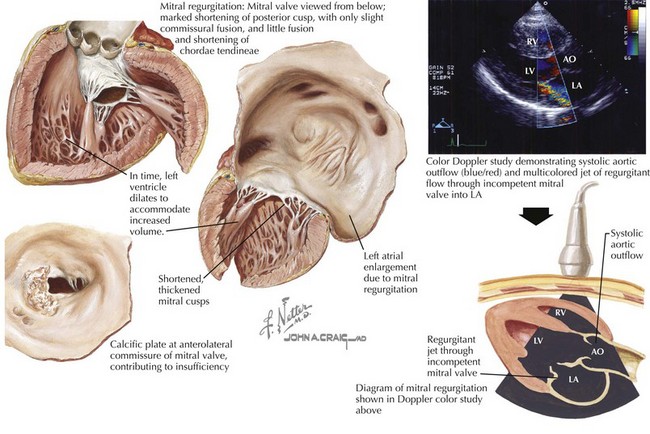


Infectious endocarditis, spontaneous rupture of chordae tendineae, or ischemic injury of a papillary muscle may cause acute loss of mitral valve integrity and acute mitral regurgitation. In these cases of abruptly increased regurgitant flow, because there is no adaptation of the left atrium or pulmonary vasculature to the increased regurgitant volumes, acute pulmonary edema may suddenly occur. Aggressive use of vasodilators is the emergent treatment, but survival usually depends on emergency repair or replacement of the valve.


Clinical Presentation


Mitral Stenosis


Patients notice the effects of moderate (valve area = 1–2 cm2) mitral stenosis with activity. With severe stenosis, dyspnea with minimal exertion and paroxysmal nocturnal dyspnea may occur. In some cases a sudden, dramatic onset of atrial fibrillation produces the first symptoms, occasionally resulting in fatal pulmonary edema. When the development of atrial fibrillation is clinically silent, the initial event may be a stroke or other thromboembolic event. The classic presentation of severe cor pulmonale with ascites and edema is rarely seen today except in medically underserved populations. Mitral valve disease increases the risk for bacterial endocarditis, which should always be considered when symptoms worsen in a previously stable patient with mitral valve disease.


Auscultatory findings in a patient with mitral stenosis include a loud first heart sound, an opening snap after the second heart sound, and a low-pitched diastolic murmur with presystolic accentuation if the patient is in sinus rhythm. The opening snap is the sound generated by sudden full opening of the mitral valve. It reflects the severity of the pressure gradient across the mitral valve, because greater LA pressures generate earlier opening than do lesser ones. Therefore, the shorter the interval from second heart sound to opening snap, the greater the pressure gradient, and the more severe the stenosis.


The characteristic diastolic, low-frequency “rumble” or murmur associated with mitral stenosis is best heard at the apex with the patient in the left lateral decubitus position and the bell over the point of maximal ventricular intensity. The rumble occurs throughout diastole, with accentuation in late diastole (presystole) in patients who have preserved normal sinus rhythm. This murmur can be difficult to hear, and is soft and brief when the stenosis is minor. Therefore, heightened awareness of possible mitral stenosis is necessary. If the murmur is inaudible, it can be accentuated by having the patient exercise before auscultation or perform maneuvers such as isometric handgrip. This murmur sequence—loud first sound, opening snap, and diastolic rumble—is specific for mitral stenosis. Murmurs that mimic mitral stenosis include the Austin Flint murmur with aortic regurgitation, mitral diastolic murmurs in patients with large intracardiac shunts, and occasionally murmurs that are caused by an LA myxoma. However, none have all three components of classic mitral stenosis.

Stay updated, free articles. Join our Telegram channel

Jun 12, 2016 | Posted by in CARDIOLOGY | Comments Off on Mitral Valve Disease

Full access? Get Clinical Tree

Get Clinical Tree app for offline access