Minor Amputations


Minor Amputations


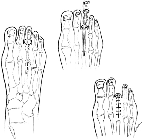
Malachi G. Sheahan, Claudie M. Sheahan and Frank B. Pomposelli


The first use of limb amputation was as an implement of punishment or torture. Hippocrates is credited with performing the first amputation for therapeutic purposes. Well into the 20th century, toe or forefoot gangrene was associated with limb loss. Leland McKittrick’s publication of his technique for transmetatarsal amputation (TMA) in 1949 led to thousands of spared limbs. For decades, however, a TMA was much more likely to succeed when infection was the indication rather than ischemia. As recently as the 1970s, up to 70% of patients who underwent minor amputation progressed to limb loss within 3 years. Better understanding of microbiology, risk modification of risk factors, and the ability to perform femoral-to-popliteal and more distal arterial bypass grafts and, more recently, catheter-based interventions, have led to contemporary limb-salvage rates approximating 80% at 5 years.


In the United States, hospital admissions for foot ulceration increase yearly. One of the most important and overlooked aspects of successful limb salvage in these patients is a properly timed and executed minor amputation. Currently, minor amputations are performed twice as frequently as major amputations. Avoidance of major amputation is critical for a myriad of reasons. Transtibial and transfemoral amputations result in inefficient ambulation. The increase in metabolic cost after these procedures is proportional to the number of functional joints that are lost. Increased oxygen consumption leads to reduced metabolic reserve and a dramatically altered exercise capacity.


Adverse consequences of major amputation are decline in quality of life, difficulty maintaining independence, and high medical costs. Body image issues, depression, and shortened life expectancy are other sequelae. The risk of amputation is approximately three times higher after age 80 years, which is precisely the population most at risk for loss of independence.


Patients with diabetes mellitus are especially prone to foot ulcers due the presence of polyneuropathy. Their predisposition for both ischemia and multimicrobial infection can result in extensive tissue loss and/or osteomyelitis and result in major limb amputation when ulcers are left unhealed.


A properly performed toe, ray, or transmetarsal amputation results in closure of the skin envelope and salvage of a walking foot. When a débridement, drainage procedure, or open amputation is required to control infection, considerable ingenuity may be needed to fashion an amputation that both closes the wound and preserves a walking foot.


Recognizing the presence of ischemia and correcting it by either bypass or catheter-based intervention is critical to achieving success with partial foot amputation. In general, any patient without palpable foot pulses should undergo evaluation for ischemia. When limb-threatening ischemia of the foot is identified, the goal of treatment should be the restoration of a palpable foot pulse when possible. In our experience, a palpable foot pulse is the most reliable clinical indicator of adequate arterial perfusion for successful healing of an amputation in the foot. If an incision and drainage procedure or open amputation is necessary to control infection in an ischemic patient, vascular evaluation should occur soon after and revascularization should be performed with 2 to 4 days in most patients to prevent further necrosis or tissue loss.


Indications


The goals of minor amputation surgery are to remove anatomic pressure points and prevent recurrent ulceration, control osteomyelitis and infection, preserve and restore the foot to optimum ambulatory function, and control and relieve pain.


A simple toe amputation is indicated in the case of irreversible tissue loss or osteomyelitis of one toe distal to the proximal interphalangeal joint. The more extensive ray amputation is performed when gangrene or osteomyelitis extends to the web space, metatarsophalangeal joint, or metatarsal head. Other indications for ray amputation include the need to decompress one deep fascial compartment or extension of skin necrosis past margins acceptable for a simple amputation.


The basic indication for transmetatarsal foot amputation is irreversible tissue loss or infection extending beyond the boundaries of a single-ray amputation but still allowing the creation of a viable plantar flap. Many patients undergoing TMA have had previous ipsilateral foot procedures such as ray amputations and incision and drainage. Common scenarios include failure of a previous toe amputation, the need for multiple ray amputations, forefoot infection involving multiple deep fascial compartments, forefoot deformity causing severe disability, and intractable pain. Because a TMA spares the tibialis anterior, peroneus longus, and peroneus brevis tendon insertions, it is a much more functional amputation than the Lisfranc, Chopart, or Syme amputations. These latter three, in our opinion, provide little to no advantage over a below-knee amputation, especially in diabetic patients.


Relative contraindications to a minor forefoot amputation include extensive tissue loss especially on the plantar aspect of the foot or the heel, untreatable foot ischemia, knee contractures, and immobility without chance for rehabilitation. These conditions usually necessitate a major amputation.


Surgical Technique


Transphalangeal Toe Amputation


The foot and leg are prepped and draped to the knee using sterile technique. The incision is made at the level of the mid-proximal phalanx down to the level of the bone. The incision should be circumferential rather than fish-mouth when possible to avoid compromising blood supply to the flap. Trimming the phalynx back to allow tension-free closure is an important principle. In the case of the first toe, the approach may be altered to include a plantar flap if possible (Figure 1).


The periosteum is stripped with an elevator. The bone is then transected with an oscillating saw at the proximal phalangeal level, approximately 1 cm proximal to the level of the skin incision. In first-toe amputations, the metatarsophalangeal joint should be spared if possible to improve gait stability. The tendons are pulled down, sharply divided, and allowed to retract. After hemostasis is obtained and the wound is irrigated, the skin is closed without tension in an anteroposterior orientation. Interrupted monofilament sutures are preferred. Dry gauze dressing is applied and full weight bearing is avoided until the wound has healed.


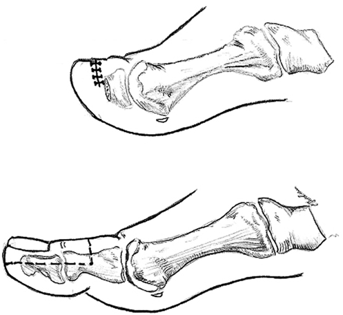
Transmetatarsal (Ray) Toe Amputation


The foot and leg are prepped to the thigh and draped using sterile technique. A racquet-type longitudinal incision is made on the dorsum of the foot parallel to the metatarsal bone extending on each side of the toe as a semielliptical incision converging on the plantar surface of the foot (Figure 2). A circular incision should be avoided if primary closure is to be attempted, because there is less tension in the closure of an elliptical incision and therefore greater chance of wound healing. A ratio of incision length to width of at least 3:1 will aid primary closure. If ulceration is present on the plantar surface at the level of the metatarsal head, this may be included in the excision as well. Preoperative assurance of adequate circulation is critical to the healing of this amputation type.


Stay updated, free articles. Join our Telegram channel

Aug 25, 2016 | Posted by in CARDIOLOGY | Comments Off on Minor Amputations

Full access? Get Clinical Tree

Get Clinical Tree app for offline access