Author
Year
Patients
Endoscope
Indication
Ash (M)
1974
11
Micro
Pleural effusion diagnosis
d’Alessandro (S)
1997
2
Micro
Pleural disease diagnosis
Nezu (S)
1997
34
Micro
Video assistance in pneumothorax surgery
Malthaner (S)
1998
3
Mini
Pleural effusion diagnosis
Nakamoto (S)
1998
31
Micro/mini
Video assistance in preoperative diagnosis
Yim (S)
2000
38
Micro
Sympathectomy for palmar hyperhidrosis
Yoon (S)
2000
130
Mini
Video assistance in pneumothorax surgery
Lazopoulos (S)
2002
54
Micro
Pleural and lung disease diagnosis
Lin (S)
2002
102
Micro
Sympathectomy for palmar hyperhidrosis
Ikeda (S)
2003
35
Mini
Video assistance in partial lung resection
Chen (S)
2003
28
Mini
Video assistance in pneumothorax surgery
Tassi (M)
2003
30
Mini
Pleural effusion diagnosis
Janssen (M)
2003
9
Micro/mini
Pleural effusion diagnosis
Chen (S)
2006
142
Mini
Video assistance in pneumothorax surgery
Sihoe (S)
2007
31
Mini
Sympathectomy for palmar hyperhidrosis
Kim (S)
2008
65
Micro
Video assistance for lung biopsies
25.2.1 Experience
Small-calibre thoracoscopes were first proposed more than 30 years ago (Ash and Manfredi 1974); however, due to the limited technology of the instrumentation, there was minimal uptake of the procedure. The idea resurfaced again in the 1990s when some thoracic surgeons used small-calibre instruments both for diagnostic (d’Alessandro 1997; Malthaner and Inculet 1998; Nakamoto et al. 1998) and therapeutic purposes (Nezu et al. 1997). There was confusion regarding the precise terminology, since some called the technique “microthoracoscopy” (d’Alessandro 1997) and others called it “minithoracoscopy” (Malthaner and Inculet 1998) or “mini thoracoscopy” or “mini-VAT” (video-assisted thoracoscopy using a miniaturized endoscope) (Nakamoto et al. 1998).
Even in the subsequent decade, experience has remained limited and predominantly surgical (Yim et al. 2000; Yoon et al. 2000; Lazopoulos et al. 2002; Ikeda et al. 2003; Chen et al. 2003; Lin and Chou 2004; Sihoe et al. 2007; Kim et al. 2008), although there have been some medical applications (Tassi and Marchetti 2003; Janssen et al. 2003). The terminology has expanded to include “needle video-thoracoscopic surgery” (NVTS) (Chen et al. 2003) or “needlescopic VATS” (Sihoe et al. 2007).
Some therapeutic applications have now become standard practice, such as for palmar hyperhidrosis (Yim et al. 2000; Lin and Chou 2004; Sihoe et al. 2007) and pneumothorax (Yoon et al. 2000; Chen et al. 2003, 2006).
The current standard treatment for palmar hyperhidrosis is thoracoscopic sympathotomy or sympathectomy, making thoracotomy obsolete. In this type of intervention, the use of small-calibre instruments became commonplace. This is because the small surgical window did necessary the use of miniendoscopes with a narrow field of vision.
In the treatment of pneumothorax, the use of small-calibre endoscopes has become more common, both in the examination of the pleural cavity and video assistance for bullectomy (Yoon et al. 2000) but also for treatment in general. Chen stated (Chen et al. 2003) that “…needlescopic VATS can be a satisfactory alternative to conventional VATS in treating primary spontaneous pneumothorax…” given that the results and incidence of recurrence were comparable, and moreover needlescopic VATS gave better cosmetic results and less residual chest pain. He also suggested the instillation of a pleurodesis agent such as minocycline to reduce recurrence (Chen et al. 2006).
Thoracoscopy with small-calibre endoscopes has also been used in various other applications such as video assistance in stapler lung biopsies for interstitial lung disease and lung nodules (Kim et al. 2008). It has also been applied for the management of traumatic pneumothorax, pericardial windows, and minor intrapleural bleeding, all of which are historically treated with a large-calibre thoracoscope (Lazopoulos et al. 2002).
In the field of medical thoracoscopy, the experience with small-calibre endoscopes is more limited. In 2003, we published our initial case study (Tassi and Marchetti 2003) using a 3 mm thoracoscope for diagnostic thoracoscopy in pleural effusions. This technique allowed for pleural biopsies and a diagnostic yield of 93.4 %, which was similar to that of standard thoracoscopy. Janssen (Janssen et al. 2003) compared the results obtained from a 7 mm thoracoscope with those from two types of small-calibre endoscopes (3.5 and 2 mm). The diagnostic yield from the 7 and 3.5 mm instruments was 100 %, whereas the yield from the 2 mm endoscope was 40 %.
25.2.2 Instruments
As noted above, small-calibre endoscopes are defined as miniendoscopes if their diameter is 2–5 mm (Fig. 25.1) and microendoscopes or needlescopes if the diameter is 2 mm or less (Tu and Advincula 2008).
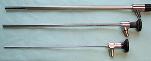
Fig. 25.1
From top to bottom, 10, 5, and 3 mm 0° telescopes
Essential requirements to carry out an adequate thoracoscopy include acceptable illumination of the cavity to be examined and sufficiently high definition images. These aspects are directly linked to the size of the instrument, and if inadequate, then examination and identification become difficult.
With minithoracoscopes, as with larger-calibre traditional thoracoscopes, light is transmitted by means of the classical Hopkins rod-lens system which gives good visibility and sufficient depth of light penetration. Although the quality is slightly inferior to that obtained by standard thoracoscopy, it is sufficient for the examination of the pleural cavity and for performing adequate biopsies especially when taken with forceps of ~3 mm (Fig. 25.2).
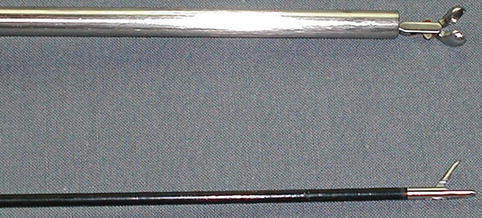
Fig. 25.2
Seven millimeter (above) and 3 mm (below) forceps with the same 7 mm opening
With microendoscopes and needlescopes light is generally transmitted through fibre-optic bundles, though recent developments have seen needlescopes produced which use the Hopkins system (Figs. 25.3 and 25.4).

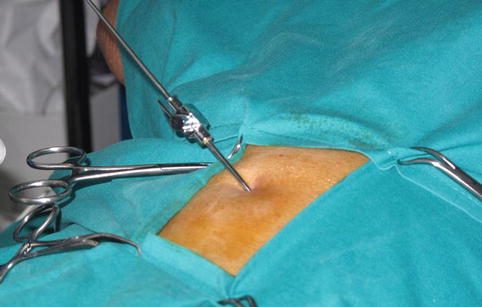
Fig. 25.3
Typical instruments used in needlescopic thoracoscopy: 2 mm forceps (above) and 2 mm needle thoracoscope (below)
Fig. 25.4
Two millimeter thoracoscope introduced into the pleural cavity through a Boutin thoracocentesis needle
Although the visibility is adequate, the definition is not always optimal. Due to the small calibre, the temperature of the instrument can change quickly with each introduction and extraction, thus subjecting the optic to frequent misting. In addition, since the optic and accessory instruments are flexible, they can at times oscillate and make it more difficult to control a manoeuvre which requires precision.
Biopsies taken with forceps of ~2 mm are often inadequate because of their small size.
A large variety of accessory instruments is now available both for minithoracoscopy and for micro/needlethoracoscopy: forceps, scissors, blunt probe, biopsy punch, grasps, aspiration/irrigation cannulae, etc. All these instruments, together with small endoscopes, are very fragile and should be handled with great care. It should be stressed that their use requires both ability and experience in standard thoracoscopy. The technological advances which have enabled the creation of these instruments could further improve their diagnostic sensitivity and, in turn, extend their application.
25.3 Minithoracoscopy
Minithoracoscopy is currently defined as endoscopy using small instruments with a diameter from 2 mm up to 5 mm. It is more appropriate in medical thoracoscopy, since it permits good visualization of the pleural cavity and if required, biopsy.
In our opinion, minithoracoscopy was never considered to be a substitute for traditional medical thoracoscopy, which, over the years, has demonstrated efficacy and reliability and, in addition, has dedicated optics, forceps, and accessory instruments. It was considered to be a complementary diagnostic approach, which is minimally invasive, safe, and cost-effective and, in certain cases, may be seen as a substitute for the traditional intervention.
Stay updated, free articles. Join our Telegram channel

Full access? Get Clinical Tree


