Percutaneous closure of an atrial septal defect has been increasingly used, and complications have been rare. We report the case of a 63-year-old man who had undergone endovascular closure of a secundum atrial septal defect months earlier. The occluder was later found in the abdominal aorta.
Case Report
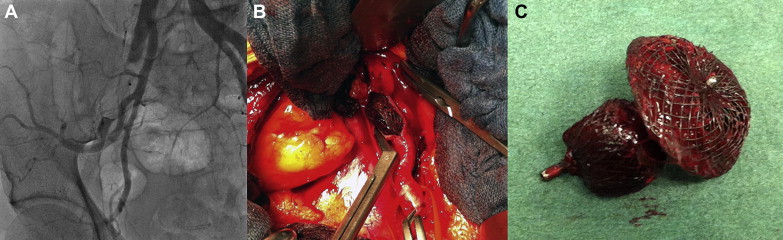
In December 2012, a 63-year-old man was referred with acute right leg ischemia due to migration of the atrial septal occluder into the right external iliac artery. He had arterial hypertension and had experienced a cryptogenic stroke on October 6, 2012 that was successfully treated with thrombolysis therapy. After that episode, transthoracic echocardiography revealed a secundum atrial septal defect (ASD) with a maximum diameter of 11.5 mm and a pulmonary flow to systemic flow ratio of about 1.4. On October 23, 2012, a 12-mm AMPLATZER septal occluder (ASO) device (St. Jude Medical, North Plymouth, Minnesota) was placed through left percutaneous femoral vein access, with the patient under local anesthesia. The patient was discharged on the second day after the procedure in good condition. The first transthoracic echocardiographic follow-up study showed ASD patency with migration of the ASO. Three days after the transthoracic echocardiographic scan, the patient underwent catheterization, which demonstrated the presence of the ASO in the abdominal aorta. Several unsuccessful attempts were performed to capture and remove the ASO using an AMPLATZER gooseneck snare kit. It was only possible to move the ASO as far as the right external iliac artery ( Figure 1 ). Because of the impossibility of ASO endovascular removal and the onset of right limb ischemia, the patient was sent to our center. After a duplex scan confirmed the presence of the ASO in the right external iliac artery, the ASO was surgically removed, with restoration of the normal blood flow. The patient was discharged on the fourth postoperative day in good general condition.
Discussion
Of all the possible complications, device embolization is 1 of the most common, with an incidence of 0.4% to 1.1%. ASO dislodgement can occur if a size discrepancy in the defect or excess space is present between the ASD and ASO. Several reasons can explain this discrepancy. First, ASDs rarely have a perfect circular shape; therefore, it could be difficult to accurately measure the largest diameter of the defect. Second, the flexibility and redundancy of the tissue surrounding the defect could result in a larger defect when the defect itself is stretched with the balloon and in the presence of a floppy rim. Percutaneous foreign body retrieval is a well-accepted technique to remove migrated devices, because it obviates major cardiovascular surgery, with high efficacy and few complications. In the published data, the most common sites of embolization have been the cardiac chambers, pulmonary artery, and aortic arch. Reviewing the English language reports, we found only 5 case reports of ASO migration into the abdominal aorta and only 1 into the iliac artery, all without limb ischemia. In 2 cases, after surgical exposure of the common femoral artery, a sheath (18F and 20F, respectively) was positioned close to the ASO device. It was grabbed and pulled partially inside the sheath and then down to the arteriotomy site. In 1 case, owing to total incorporation of the device, endoluminal retrieval was not possible; thus, the device was surgically removed through a medial laparotomy approach. In 2 cases, the ASO device was retrieved from the descending aorta by making a horizontal cut in the ascending aorta. In another case, the ASO device, which had migrated to the level of the aortic bifurcation, was removed by laparoscopic extraction. Our report is the first of a case of the ASO device that had dislocated into abdominal aorta and, after an unsuccessful endovascular attempt, had embolized into the iliac vessel, resulting in the complication of an ischemic limb.
Stay updated, free articles. Join our Telegram channel

Full access? Get Clinical Tree


