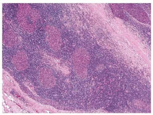
Micronodular Thymoma with Lymphoid Stroma
Borislav A. Alexiev, M.D.
Anja C. Roden, M.D.
Terminology
Micronodular thymoma (MNT) is an organotypical thymic epithelial neoplasm characterized by multiple, discrete epithelial nodules separated by an abundant lymphocytic stroma that usually contains prominent germinal centers.1 The epithelial component is similar to type A thymoma.
Incidence and Clinical
MNT is an uncommon entity and corresponds to1% to 5% of all thymomas.1 The age at manifestation ranges from 45 to 95 years. There is no sex predilection.1
MNT is rarely (<5%) associated with paraneoplastic myasthenia gravis. Other autoimmune phenomena that are common in other thymoma types have not been reported. Clinical features usually are related to the size and local extension of the tumor. MNT can be asymptomatic and is found incidentally upon x-ray, CT, or MRI examination.1
Gross Pathology
Grossly, MNT is usually encapsulated, and the cut surface shows multiple tan-colored nodules of various sizes separated by white fibrous bands and cystic changes.
Microscopic Pathology
Histologically, MNT shows a micronodular growth pattern.1,2,3,4,5,6,7 Multiple, discrete epithelial nodules are separated by an abundant lymphocytic stroma that may contain lymphoid follicles with prominent germinal centers (Fig. 104.1). There are a variable number of mature plasma cells. The epithelial nodules are composed of slender or plump spindle cells with bland-looking oval nuclei and inconspicuous nucleoli (Fig. 104.2). Micro- and macrocystic areas, particularly in subcapsular localization, are common. There are no Hassall corpuscles or perivascular spaces. Mitotic activity is
absent or minimal. The epithelial nodules contain few interspersed lymphocytes.
absent or minimal. The epithelial nodules contain few interspersed lymphocytes.
Stay updated, free articles. Join our Telegram channel

Full access? Get Clinical Tree


