Meningothelial-Like Nodule
Allen P. Burke, M.D.
Marie-Christine Aubry, M.D.
General
Meningothelial-like nodules (MLNs) have several designations, including “minute meningothelial-like nodule,” “meningothelial nodules,” and “chemodectoma.” They are quite common as incidental findings and only rarely cause pulmonary symptoms, when diffuse and bilateral in so-called diffuse pulmonary meningotheliomatosis.1 Although they were initially thought to be neuroendocrine and therefore called chemodectoma, subsequent studies confirmed the meningothelial nature of the cells of MLN.2
Histogenesis
The cells of MLN share many features with cells of meningiomas, including histologic appearance, ultrastructural finding, and frequent immunohistochemical expression of epithelial membrane antigen and CD56. MLNs have not been reported in children; hence, they are not thought to be congenital. A loss of heterozygosity (LOH) study comparing single MLN to meningiomas found multiple events only in meningiomas. The same study demonstrated that cases of multiple MLNs (meningotheliomatosis) occasionally harbored multiple LOH events, but at a lower rate than meningiomas, suggesting that single MLNs are reactive and that multiple tumors may represent the transition between a reactive and neoplastic proliferation.3
Incidence and Clinical Presentation
Meningothelial nodules are common, and many are likely overlooked. The incidence in lung resections for other conditions ranges from 7% to 42%.4,5 They are twice as common in women as men, without predilection for any lobe. They are increased in frequency in resections for malignancies, especially adenocarcinomas, as opposed to benign conditions,4 and are also common in lung resections showing infarcts, thromboembolic disease, and respiratory bronchiolitis.5 They are multiple in over 50% of cases.5 In rare patients, MLN may be so numerous as to mimic an interstitial lung disease or metastatic malignancy. Indeed, these patients present with shortness of breath with mild restriction on pulmonary function tests and bilateral reticulonodular infiltrates radiologically. This disease manifestation has been called diffuse pulmonary meningotheliomatosis.1
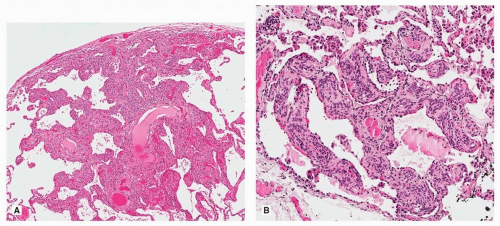
 FIGURE 70.1 ▲ Meningothelial-like nodule. A. Low magnification showing irregular-shaped nodule with the tumor cells insinuated within alveolar septa. The appearance is often not that of a discrete solid nodule but rather nodular interstitial thickening by the tumor cells. B. Another example demonstrating the interstitial growth pattern of most MLN.
Stay updated, free articles. Join our Telegram channel
Full access? Get Clinical Tree
 Get Clinical Tree app for offline access

|