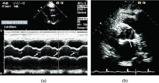
Figure 40.2 M-mode echocardiographic images illustrate the eccentric closing line (arrows) of aortic valve (a). Aortic valvular short-axis view indicates the opening of bicuspid valve; it is like a fish’s mouth (b, arrow) in the same patient with membranous ventricular septal defect.
He underwent an elective closure of membranous VSD without complications.
Discussion
Stay updated, free articles. Join our Telegram channel

Full access? Get Clinical Tree


