Fig 1.
Surfactant deficiency. A 27-week-gestation infant on the first day of life. Portable chest radiograph shows hypoinflated lungs with scattered air-bronchograms
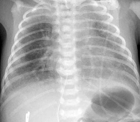
Fig 2.
Asymmetric surfactant administration. A 28-week-gestation infant on the first day of life. Chest radiograph obtained after in-tratracheal surfactant administration shows asymmetric central inflation of the right mid-lung due to asymmetric surfactant administration to the right lung
Pneumonia
The incidence of pneumonia in the newborn appears to be inversely proportional to gestational age, occurring in up to 4% of premature infants <1000 g. The most common pathogens in this age group currently include Escherichia coli, group B streptococcus, and staphylococcus species. Although alveolar infiltrates may be seen in the premature infant with pulmonary infection (Fig. 3), both the clinical signs and the radiographic features of pneumonia may be indistinguishable from those of hyaline membrane disease.
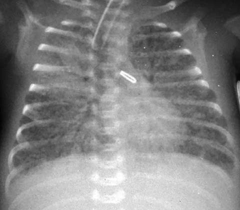
Fig 3.
Neonatal pneumonia. A 26-week-gestation infant at 2 weeks of life. Portable chest radiograph shows a focal right upper lobe pneumonia with air bronchograms superimposed on early chronic lung disease
Transient Tachypnea of the Newborn
Transient tachypnea of the newborn is one of the most common causes of respiratory distress in the older premature and full-term infant. The basic underlying patho-physiology is one of delayed clearing of fetal lung fluid. Risk factors include prematurity and birth by cesarean section, usually without preceding labor. Respiratory difficulties begin within a few hours of birth, lasting between a few hours up to 2 days in uncomplicated cases. Chest radiographs typically show increased perihilar bronchovascular markings and fluid in the pulmonary fissures (Fig. 4). The lungs may vary in their degree of inflation, from normal to hyperinflated.
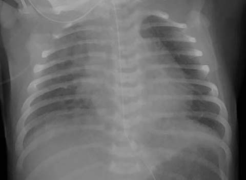
Fig 4.
Transient tachypnea of the newborn. A 36-week-gestation infant, born by cesarean section, on the first day of life. Chest radio-graph shows increased central bronchovascular markings and a mild interstitial prominence
Meconium Aspiration
Meconium aspiration syndrome consists of fetal acidosis and subsequent hyperperistalsis, passage of meconium, fetal gasping, and aspiration of meconium into the trachea and lungs [2]. Thick meconium may cause physical obstruction of the distal airways, resulting in patchy, alternating areas of pulmonary overinflation and atelectasis. Meconium also inactivates surfactant and causes secondary surfactant deficiency and poor lung compliance. The clinical course of many patients with meconium aspiration syndrome is complicated by pulmonary hypertension, which increases illness severity and worsens clinical outcomes. The radiographic features of meconium aspiration vary widely, depending on the severity of aspiration. The mildest cases manifest as pulmonary hyperinflation with scattered interstitial pulmonary opaci ties, while in more severe cases patchy alveolar and interstitial infiltrates and more pronounced hyperinflation are seen, often together with pneumothorax and pneumomediastinum as complications (Fig. 5).
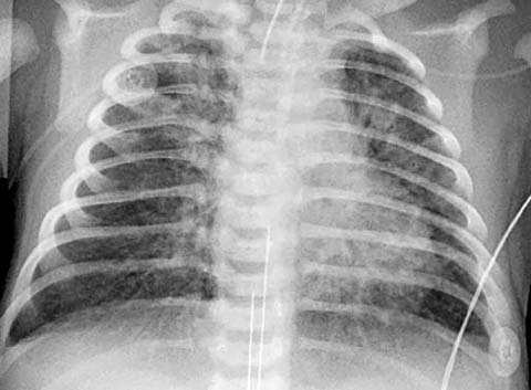
Fig 5.
Meconium aspiration. A 39-week-gestation infant on the first day of life. Heavy meconium staining and low APGAR scores at birth were recorded. Portable chest X-ray shows hyperinflated lungs with coarse bilateral interstitial opacities and focal right upper lobe atelectasis
Complications of Respiratory Support
Pulmonary air leaks are complications of greatest concern in newborns receiving positive pressure ventilation. They can manifest as pulmonary interstitial emphysema, pneumomediastinum, pneumothorax, and intravascular gas. As alveoli become overdistended and rupture, gas is forced into the connective tissues of the lung and can dissect along the interlobular septae toward the hilum of the lung. This pattern is of flame-shaped lucencies that may be bilateral or unilateral and is characteristic of pulmonary interstitial emphysema (PIE). Increasing distention can progress to plural or mediastinal air collections with or without PIE. In rare cases, gas is forced through lymphatic channels into the vascular tree, resulting in cardiovascular collapse (Fig. 6).
Stay updated, free articles. Join our Telegram channel

Full access? Get Clinical Tree


