Management Implications of Massive Left Ventricular Hypertrophy in Hypertrophic Cardiomyopathy Significantly Underestimated by Echocardiography but Identified by Cardiovascular Magnetic Resonance


Cardiovascular magnetic resonance (CMR) is a high spatial resolution, 3-dimensional tomographic imaging technique which may identify regions of massive left ventricular hypertrophy (particularly when confined to the anterolateral free wall) in which the extent of wall thickness is underestimated with traditional 2-dimensional echocardiography in patients with hypertrophic cardiomyopathy (HC). This observation may have potential implications on management strategies as extreme left ventricular hypertrophy is a primary risk factor for sudden death in HC and therefore supports an expanding role for CMR in the evaluation of HC patients.


Case Descriptions


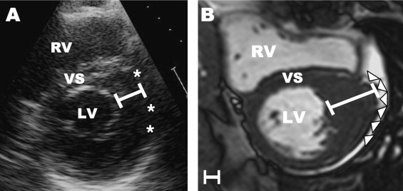
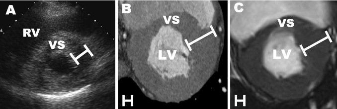
The cardiovascular magnetic resonance (CMR) images presented herein ( Figures 1 to 3 ) expand the role of advanced imaging techniques for hypertrophic cardiomyopathy (HC). CMR (and computed tomography) identified massive degrees of left ventricular (LV) wall thickening (≥30 mm), confined to the anterolateral free wall and substantially underestimated in magnitude by cross-sectional 2-dimensional echocardiography.


Figure 1


Massive left ventricular hypertrophy identified only by CMR and significantly underestimated by 2-dimensional echocardiography in HC. A 46-year-old man with HC was evaluated for atypical chest pain following a normal myocardial exercise perfusion study. (A) Two-dimensional echocardiographic end-diastolic basal short-axis view demonstrates a maximal left ventricular wall thickness of 18 mm in the anterolateral free wall, consistent with the diagnosis of HC. (B) In the same patient, end-diastolic short-axis CMR at the same level of the left ventricle (LV) shows a focal area of massive left ventricular hypertrophy (35 mm) in the same region of the left ventricular wall reported to be 18 mm by echocardiography. This segmental pattern of wall thickening was not appreciated by echocardiography because of the limited spatial resolution of the lateral left ventricular wall (asterisks) and the expectation that the epicardial border of the lateral wall would conform to a spherical shape. The finding of massive hypertrophy on CMR characterized this patient as at high risk and prompted recommendation for implantable cardioverter-defibrillator therapy for the primary prevention of sudden death. Calibration markers are 1 cm apart. RV = right ventricle; VS = ventricular septum.


Figure 2


Massive left ventricular hypertrophy recognized only by cardiac computed tomography (CT) and CMR. A 35-year-old man with HC was evaluated for atypical chest pain. (A) Two-dimensional echocardiographic end-diastolic basal short-axis image demonstrates diffuse hypertrophy with a maximum left ventricular wall thickness of 19 mm in the anterolateral wall. (B) Cardiac CT demonstrated a maximum wall thickness of 31 mm in the anterolateral free wall. (C) End-diastolic short-axis CMR image shows wall thickness measurement of 31 mm, identical to CT at the same level of the left ventricle (LV). Calibration markers are 1 cm apart. RV = right ventricle; VS = ventricular septum.

Only gold members can continue reading. Log In or Register to continue

Stay updated, free articles. Join our Telegram channel

Dec 23, 2016 | Posted by in CARDIOLOGY | Comments Off on Management Implications of Massive Left Ventricular Hypertrophy in Hypertrophic Cardiomyopathy Significantly Underestimated by Echocardiography but Identified by Cardiovascular Magnetic Resonance

Full access? Get Clinical Tree

Get Clinical Tree app for offline access