Magnetic Resonance Arteriography for Assessment of Infrainguinal Occlusive Disease


Magnetic Resonance Arteriography for Assessment of Infrainguinal Occlusive Disease


Nyali E. Taylor and Jeffrey P. Carpenter


Diagnostic imaging has changed dramatically since the 1960s and the advent of conventional arteriography, particularly for peripheral vasculature. A major drawback of conventional arteriography is a failure to visualize infrageniculate arteries as well as other modalities, such as magnetic resonance arteriography (MRA).


Historically, up to 70% of preoperative conventional arteriography evaluations failed to enhance patent distal vessels. MRA was able to identify 24% of additional vessel segments when compared to conventional arteriography in our original series. Although intraarterial digital subtraction angiography (DSA) has supplanted conventional arteriography, recent literature continues to confirm DSA’s inferiority at opacifying pedal vessels when compared to MRA. For instance, Andriesek found that DSA missed 35% of patent pedal vessels in diabetics when compared with a newer MRA adjunctive imaging modality.


The demonstration of patency of a distal vessel by conventional arteriography necessitates that a bolus of proximally injected contrast medium traverse multiple segmental occlusions before it can opacify the patent downstream vessels. In the process, there are multiple dilutions and reconstitutions of contrast medium as it crosses arteriolar and capillary beds through circuitous collaterals to the distal circulation. In patients with limb-threatening ischemia and severe occlusive disease, opacification of small distal vessels presents a great challenge to contrast-based imaging techniques. The data are not as clear-cut at more proximal stations, but MRA may be emerging as the modality of choice for diagnostic imaging of pedal vasculature.


Fundamental Principles of Magnetic Resonance Arteriography


MRA works on the principle of differences in proton emission detected as a signal. This is accomplished by placing a patient within a large external magnetic field (1.5–3.0 Tesla) to align the patient’s protons in parallel with the field. Magnetic field gradients work to alter the protons’ resonance frequencies, and a resonant coil tuned to the appropriate frequency generates the radiofrequency signal that is detected by a receiver coil. Pulse sequences are the specific combination of radiofrequency and magnetic gradients that are used to create the image. These sequences create a T1-assisted or T2-weighted image, enhancing specific tissue types (e.g., fluid, fat). MRA uses T1-weighted images. The protons emit an echo (signal) based on their characteristics and the field size. The signal is then amplified, digitized, and processed to formulate images.


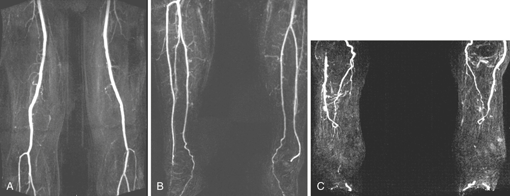
Technological advances have enabled MRA to acquire higher-resolution images in less time. Images may be obtained by noncontrast or contrast-enhanced methods. Time-of-flight (TOF) noncontrast imaging is based on signal differences between saturated and unsaturated protons in blood. This technique has limited use in the era of contrast-enhanced MRA, and it is employed when contrast is contraindicated. Contrast-enhanced MRA most often uses gadolinium, which is an element whose properties shorten the T1 relaxation, causing blood to be more visible than the surrounding tissues (Figure 1). It is designed to produce an effect on the protons in the surrounding water. Thus, minute amounts of MRA contrast may be detected by its effect on multiple water molecules, whereas an equivalent dose of iodinated contrast is not detectable directly by conventional arteriography or computed tomography angiography (CTA). Contrast agents with different properties are under investigation to determine if they improve image quality, sensitivity, or specificity of MRA.


Clinical Experience with Magnetic Resonance Arteriography


In 1992, we published an initial experience with MRA. At that time, employing two-dimensional TOF techniques, we found that 52% of MRA and conventional arteriography studies had exact agreement in patients with severe, symptomatic peripheral arterial disease. Of the remaining vessel segments, MRA identified 24% of those not seen by conventional arteriography. This increased detection of patent runoff vessels was most pronounced in the distal arterial segments. MRA maintains certain advantages over conventional arteriography and DSA.


MRA, compared to CTA, has the advantage of not being plagued by artifactual interpretation of occlusion in the presence of severe calcification. Calcium does not appear on MRA images, which is often a source of confusion on CTA images. MRA detects the presence of flow, producing its exquisite sensitivity for distinguishing stenosis from occlusion.


Both TOF- and contrast-enhanced MRA have proven to be equally accurate for diagnosis when compared to conventional or digitally subtracted angiography. In a meta-analysis of 28 prospective original studies, TOF was found to have a sensitivity and specificity of 93% and 88%, respectively while contrast-enhanced MRA was 96% for each. Three-dimensional gadolinium-enhanced techniques were found to have a diagnostic odds ratio of 2.8 for stenoses greater than 50% or occlusions, indicating the adequacy of that modality to identify disease. More than 80% of the study populations were classified as claudicants.


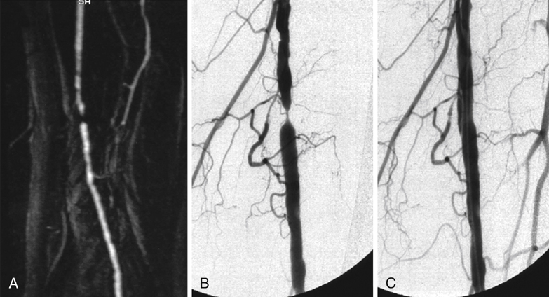
In comparison to selective DSA, contrast-enhanced MRA performs equally well in high-flow arterial segments (Figure 2). The differences become more apparent in low-flow regions, with MRA visualizing more segments in the tibial artery that were considered occluded by DSA evaluation (100% sensitivity and specificity). Overall, contast-enhanced MRA was ascertained to be comparable to DSA, with 100% and 98% accuracy, respectively. A common finding noted in the literature is that DSA or conventional arteriography overestimates occlusions, likely affecting surgical interventions and outcomes. MRA reliably detects patent runoff vessels that are occult to conventional arteriography, leading to a higher limb-salvage rate by facilitating reconstructive surgical options.


Stay updated, free articles. Join our Telegram channel

Aug 25, 2016 | Posted by in CARDIOLOGY | Comments Off on Magnetic Resonance Arteriography for Assessment of Infrainguinal Occlusive Disease

Full access? Get Clinical Tree

Get Clinical Tree app for offline access