Lymphedema and Nonoperative Management of Chronic Venous Insufficiency
Gregory L. Moneta
Lymphedema and chronic venous insufficiency (CVI) share similar principles of nonoperative management but have different pathophysiologies and operative treatments. This chapter will focus on the spectrum of management of lymphedema and nonoperative therapy for chronic venous insufficiency. Operative therapy for chronic venous insufficiency will be covered separately.
Lymphedema
Pathophysiology
Lymphedema is extremity swelling resulting from a reduction in lymphatic transport. Reductions in lymphatic transport may result from a number of anatomic or functional abnormalities, such as dermal lymphatic hypoplasia; acquired stenosis or obliteration of the axial lymphatics; or acquired or congential absence or malfunction of lymphatic valves. The functional result common to all these abnormalities is pooling of lymph within the interstitial space and swelling of the subcutaneous tissues and skin. While most lymphedema of clinical importance to vascular surgeons involves an extremity, lymphedema can affect the skin and subcutaneous tissues anywhere.
The most widely used classification of lymphedema is based on whether there is a known etiology of the lymphedema. There are primary and secondary forms of lymphedema. Primary lymphedema results from an unknown cause. It may have a genetic component of uncertain phenotypic expression. Primary lymphedema is subdivided into congenital lymphedema, lymphedema praecox, and lymphedema tarda.
Congenital lymphedema can involve a single lower extremity, multiple limbs, the genitalia, or the face. The edema is typically present at birth. Milroy’s disease is a form of congenital lymphedema generally affecting the lower extremities. It results from an absence of the dermal lymphatics. The axial lymphatics are normal. The children typically develop lower-extremity swelling shortly after birth that becomes more pronounced with attempted ambulation.
Lymphedema praecox is the most common form of primary lymphedema, accounting for 94% of cases. It is far more common in women than men, with the gender ratio favoring women 10:1. The onset of swelling is during the childhood or teenage years and involves the foot and calf. Lymphedema praecox affects primarily the axial lymphatics with varying combinations of obliteration and reflux of the axial lymphatic vessels. The onset of the lymphedema often follows an injury so trivial that it is difficult to imagine how the injury could have directly resulted in limb swelling. It seems more likely that the minor injuries associated with the onset of lymphedema praecox are circumstantial and not actually related to the onset of the disease. Lymphedema praecox has an uncertain prognosis. It often begins with foot and ankle swelling. The swelling may remain confined to the distal aspect of a single extremity or may progress more proximally. Involvement of the opposite lower extremity and even upper extremities may occur.
Lymphedema tarda is uncommon, accounting for less than 10% of cases of primary lymphedema. The pathophysiology, anatomic abnormalities, and prognosis appear similar to lymphedema praecox except that the onset of the edema is later in life than in lymphedema praecox.
Secondary lymphedema is far more common than primary lymphedema. Secondary lymphedema is a result of acquired lymphatic obstruction or disruption. Lymphedema of the arm following axillary node dissection for treatment of breast cancer is the most common cause of secondary lymphedema in the United States and other developed countries. Other causes of secondary lymphedema include trauma, radiation therapy, or malignancy. World wide, filariasis, which causes elephantiasis, is the most common cause of secondary lymphedema.
Clinical Diagnosis
The diagnosis of lymphedema is usually based on the combination of the history, physical examination, and the exclusion of other potential causes of limb swelling. Many conditions can cause edema, particularly of the lower extremities. Distinguishing lymphedema from other more common causes of leg swelling is, however, usually not difficult. If the onset of edema is bilateral, the cause of the limb swelling is likely not lymphedema secondary to an anatomic lymphatic abnormality. Bilateral pitting edema is typically associated with congestive heart failure, renal failure, or a hypoproteinemic state. The most common dilemma is to distinguish the swelling of lymphedema from that of venous insufficiency.
Patients with lymphedema and venous disease both commonly complain of fatigue and heaviness of the affected extremity. Lymphedema is usually, but not always, painless. Patients with lymphedema may complain of pain and discomfort but, in general, the pain
component of lymphedema is less than that of chronic venous insufficiency.
component of lymphedema is less than that of chronic venous insufficiency.
In patients with lymphedema, as in those with venous insufficiency, the limb circumference increases throughout the day and decreases overnight when the patient is in bed. The lymphedematous limb, however, rarely completely normalizes even with a prolonged period of bedrest and leg elevation. This is in contrast to swelling secondary to venous insufficiency where the response to bedrest and leg elevation is usually more dramatic.
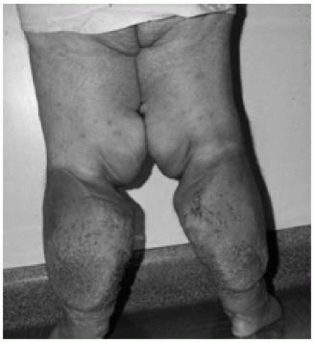
Lymphedema swelling classically involves the dorsum of the foot. Venous swelling usually does not extend onto the foot. The toes in established lymphedema have a “squaredoff” appearance. There is usually no swelling of the toes in pure venous insufficiency. In advanced lymphedema, usually neglected and inadequately treated cases, hyperkeratosis of the skin develops (Fig. 72-1) and fluid weeps from lymph-filled vesicles. Hyperpigmentation, a hallmark of long-standing venous insufficiency, is not a part of lymphedema. While ulceration can occur with lymphedema, it is very unusual.
Recurrent cellulitis is a common complication of lymphedema. Repeated infection results in further lymphatic damage, worsening existing disease and putting the patient at increased risk of future infection. The clinical presentation of cellulitis ranges from subtle erythema and worsening of edema to a rapidly progressive soft tissue infection with systemic toxicity.
Imaging Studies
Duplex Ultrasound
As noted above it is sometimes difficult to distinguish the early stages of lymphedema from venous insufficiency. Duplex ultrasound of the venous system can determine if venous reflux is present and perhaps contributing to extremity edema. Duplex is recommended to exclude venous insufficiency in patients with possible lymphedema. It may change therapy through identification of surgically correctable venous reflux.
CT Scanning/MR Imaging
CT scanning or MR imaging can be used to help exclude a pelvic process that may result in secondary lower-extremity swelling. An occasional pelvic vascular malformation or tumor is discovered. The yield is low.
CT scanning of the chest or thoracic outlet region may also discover an underlying cause of upper-extremity swelling. Primary lymphedema of an upper extremity is sufficiently uncommon that CT scanning or MR imaging of the chest and thoracic outlet regions in patients with unexplained upper-extremity swelling seems prudent.
Other Imaging Studies
Most diagnostic modalities specifically for lymphedema have limited use in routine clinical practice, although some will argue they are necessary to unequivocally establish a diagnosis of lymphedema and to uncover the rare case amenable to surgical therapy. The diagnostic modalities specifically for lymphedema are, however, relatively invasive compared to most modern vascular diagnostic techniques. They are certainly tedious and rarely change management.
Lymphoscintigraphy
Isotope lymphoscintigraphy identifies lymphatic abnormalities. A radio-labeled sulfur colloid is injected subdermally in an interdigital region of the affected limb. The lymphatic transport is monitored with a whole-body gamma camera, and major lymphatics and nodes can be visualized.
Radiologic Lymphology
Radiologic lymphology visualizes lymphatics with colored dye injected into the hand or foot. The visualized lymphatic is exposed through a small incision and cannulated. An oil-based dye is injected over several hours. The lymphatic channels and nodes are visualized with traditional roentgenograms.
Management
Management of lymphedema is primarily nonoperative and is directed toward maintaining as near-normal limb circumference as possible within the constraints imposed by the disease and necessary activities of daily living.
Education
Perhaps the most important component of the management of lymphedema is patient and family education. Patients and their families must understand there is no cure for lymphedema and that the primary goals of treatment are to minimize swelling and to prevent recurrent infections. Controlling the chronic limb swelling can improve sensations of discomfort, heaviness, and tightness and may help prevent infection. It may also potentially reduce the progression of disease.
Leg Elevation
Limb elevation is an important aspect of controlling swelling. Periodic limb elevation above the level of the heart is the first recommended intervention in patients with mild lymphedema. Several days of profound limb elevation and strict bedrest may be required in the initial management of difficult cases. Under such circumstances limb circumference can be made to dramatically decrease. However, continuous elevation throughout the day can interfere with quality of life more than lymphedema itself. Limb elevation is an important adjunct to lymphedema therapy, but it is not the mainstay of treatment.
Compression Garments
Compression garments are the foundation for treatment of lymphedema and are widely employed. No matter what other modalities are used, the patients must wear compressive garments on the involved extremity whenever they are up and about. Patients with severe lymphedema may even benefit from wearing stockings at night while sleeping.
Elastic compression stockings reduce the amount of swelling in the involved extremity by decreasing the accumulation of edema when the extremity is dependent. When worn daily, compression stockings are associated with long-term maintenance of reduced limb circumference. They offer a degree of protection against external cutaneous trauma that can precipitate cellulites. By reducing edema they may also protect the tissues against chronically elevated interstitial pressures that can lead to cutaneous thickening and hyperkeratosis.
Stay updated, free articles. Join our Telegram channel

Full access? Get Clinical Tree


