Lymphedema


Lymphedema


Thom W. Rooke and Cindy L. Felty


Lymphedema is defined as a pathologic condition in which there is an interstitial accumulation of protein-rich fluid. This condition affects more than 1 percent of the United States population and more than 150 million people worldwide. Lymphedema occurs secondary to an anatomic or functional obstruction in the lymphatics or the lymph nodes. Although it can result in edema in the truncal area, it usually occurs distally in the upper and lower extremities.


Lymphedema can be a difficult and frustrating condition to treat because most patients cannot be helped by surgery. As a result, therapy is focused around education about the disease, elastic compression, physical therapy, drugs, hygiene, and other conservative measures. Treatment is important; if patients fail to be vigilant in their treatment program, the condition typically worsens, leading to repeated infections, enlargement of the limb, thickening of the skin, disfigurement, and potential disability. Unfortunately, there continues to be a poor understanding of the theoretical and practical aspects of lymphedema treatments by many health professionals. As a result, suboptimal therapy may be offered to these patients.


Anatomy and Physiology


The lymph system is a complex network composed of fine lymphatic vessels and lymph nodes, along with other specialized structures such as spleen, thymus, and tonsils. It helps maintain water and protein balance in the tissues and assists the immune system.


Fluids, proteins, and other soluble substances extravasate from the capillaries into the interstitial space. As fluids build up, tissue pressure pushes fluid from the interstitial space into the lymph vessels. Valves located within the lymphatics ensure that the lymph flows in only one direction. The lymphatics provide a normal conduit for the return of the interstitial fluid and proteins by way of the thoracic duct back into the circulatory system. Once interstitial fluid enters the lymphatic vessels it is referred to as lymph fluid.


The rate of lymph flow can be increased by skeletal muscle contraction, increased heart rate, and passive movements from other body parts. The velocity of lymph flow can increase 10-fold over resting values during exercise.


Pathophysiology


Lymphedema develops when the lymphatic load exceeds the transport capacity of the lymphatic system. When this occurs, the tissue becomes inundated with the protein-rich edema fluid. Without treatment, the protein-rich interstitial fluid is eventually replaced by fibrous tissue and collagen, which can lead to progressive fibrosis and, in later stages, irreversible tissue enlargement.


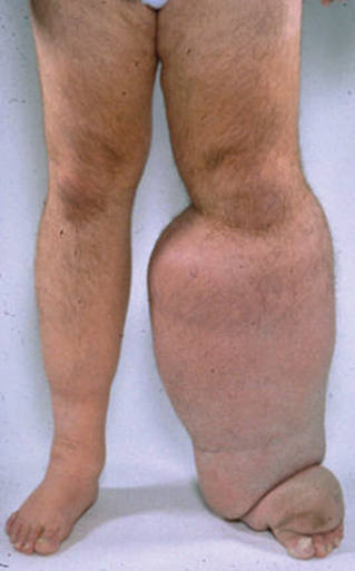
Lymphedema is often described by stages. The first stage, also called the reversible stage, is manifested by edema of a pitting nature. The edema is soft, and the swelling typically resolves overnight with simple elevation of the affected limb. If the lymphedema remains untreated, it produces a progressive hardening of the tissues, which occurs secondary to scarring and the proliferation of connective and/or adipose tissue. This stage is known as irreversible or stage II lymphedema and is manifest by edema that does not resolve with overnight elevation. At this point, the skin loses its pitting character and begins to thicken. Stage III (or lymphostatic elephantiasis) is manifested by a tremendous increase in the volume of the affected limb. The hardening of the dermal tissues and papillomas that subsequently develop can give the patient the appearance of having elephant skin (Figure 1).


Classification


Lymphedema can be characterized as primary or secondary. Primary lymphedema occurs in the absence of any obvious cause. It is more common in girls and women and usually occurs in the lower extremity. Primary lymphedema is thought to arise from an inborn defect in the development of lymphatic vessels; it can be present at birth or can occur before the age of 1 year, in which case it is classified as congenital lymphedema. Lymphedema occurring between the ages of 1 and 35 years is referred to as lymphedema praecox. Lymphedema tarda is generally the name given to primary lymphedema occurring after the age of 35 years. Congenital lymphedema occurring in a familial pattern is referred to as Milroy’s disease.


Secondary lymphedema is caused by injury, scarring, or excision of the lymph nodes. It can occur as a result of surgery in which the lymph nodes are removed, or it can develop as a side effect of radiation therapy. Secondary lymphedema is occasionally caused by trauma or by chronic infections of the lymph system. Infections of the lymph system are generally bacterial, with Streptococcus species being the most common pathogen. In some parts of the world, especially in the tropics, secondary lymphedema is typically caused by parasitic infections.

Stay updated, free articles. Join our Telegram channel

Aug 25, 2016 | Posted by in CARDIOLOGY | Comments Off on Lymphedema

Full access? Get Clinical Tree

Get Clinical Tree app for offline access