Left Heart Catheterization


Left Heart Catheterization


Andrew N. Rassi


I. Introduction.

In 1958, Dr. Mason Sones and colleagues at the Cleveland Clinic performed the first selective coronary arteriographic procedure. Since then, left heart catheterization (LHC) has become an important tool in diagnostic cardiology. More than 1.5 million cardiac catheterizations are performed yearly in the United States. Despite the advent of other imaging modalities, coronary arteriography remains the clinical gold standard for determining the presence of significant coronary artery disease (CAD).

LHC is an invasive procedure with serious potential risks. To be competent in LHC, a cardiologist-in-training must perform at least 300 catheterizations, serving as primary operator on 200. During training, the operator must be supervised by a cardiologist who is already competent in the procedure. Because there is the ability to treat a lesion with percutaneous intervention at the same time as the diagnostic angiogram, it is important to have a plan regarding how to use the information obtained.


II. INDICATIONS.

The American College of Cardiology and the American Heart Association (ACC/AHA) have categorized reasonable indications for LHC as class I, when there is consensus that LHC is indicated, or class II, when there is no consensus that LHC is indicated, but nonetheless the procedure is frequently performed (see Table 64.1).


A. Acute myocardial infarction (MI).

LHC is given a class I indication for routine use in acute MI, particularly in those who will likely undergo primary percutaneous cardiac intervention, those in cardiogenic shock or with other evidence of hemodynamic instability, or those with mechanical complications who are likely to undergo surgical repair. It can be used with the goal of performing primary angioplasty in patients with acute ST-segment elevation MI. There is evidence of the benefit of an early invasive strategy in stable patients with non—ST-segment elevation MI. In patients with non—ST-segment elevation MI, an early invasive strategy has received a class I indication in the presence of the following risk features: elevated cardiac troponins, new or presumed new ST-segment depression, heart failure (HF), depressed ventricular function, hemodynamic instability, sustained ventricular tachycardia, previous bypass surgery, and previous coronary intervention in the last 6 months. Patients with persistent pain or unresolved electrocardiographic changes after thrombolytic therapy are also class I candidates for LHC. Routine LHC soon after thrombolytic therapy in a patient who appears to have clinically reperfused is a class IIb indication.


B. Unstable angina.

LHC is given a class I indication in the patient with refractory unstable angina that cannot be controlled by medical therapy. Its role in unstable angina that can be medically controlled is controversial.


C. Chronic stable angina.

LHC is given a class I indication for purposes of revascularization in chronic stable angina for patients whose angina is poorly controlled by medicines or who are intolerant of antianginal medications.


D. Abnormal stress test.

A stress test that is positive at a low work load (6.5 metabolic equivalents of oxygen consumption) or that is classified as high risk is a class I indication for LHC. An ST depression of 2 mm, especially in multiple leads or persisting into recovery 6 minutes, an ST elevation of 2 mm in leads without Q waves, a drop in blood pressure of> 10 mm Hg with exercise, or development of ventricular tachycardia with exercise constitutes a high-risk stress test. A high-risk stress test on a concomitant imaging modality showing left ventricle dilatation, a drop in ejection
fraction of 10%, or multiple areas of ischemia is a class I indication for catheterization. These indications hold true even if the patient is asymptomatic. Positive stress tests without high-risk criteria are class II indications for LHC.


TABLE 64.1 Indications and Contraindications to Left Heart Catheterization


Indicationsaa


Contraindicationsaa


– Acute myocardial infarction


– Active bleeding


– Unstable angina


– Coagulopathy


– Chronic stable angina, uncontrolled by medications


– Acute or chronic renal failure — Active infection


– Abnormal stress test


– Severe anemia


– Ventricular arrhythmias


– Electrolyte abnormalities


– Left ventricular dysfunction — Valvular heart disease


– Inability to lie supine (i.e., decompensated heart failure)


– Preoperative coronary assessment for cardiovascular surgery


– Preoperative risk assessment for noncardiovascular surgery


– Periodic follow-up after cardiac transplantation


– Malignant hypertension


– Extensive comorbidities—risk of revascularization likely outweighs the benefit


– Patient unable to cooperate or does not desire procedure


a See text for full details.


E. Ventricular arrhythmia.

A history of sustained polymorphic ventricular tachycardia, without obvious metabolic cause, is considered a class I indication for LHC.


F. Left ventricular dysfunction.

LHC can provide an estimate of left ventricular function and regional wall motion. Left ventricular dysfunction of unknown cause, with an ejection fraction of < 40%, is a class I indication for LHC to rule out CAD.


G. Valvular heart disease.

LHC can be performed to assess the severity of outflow tract obstruction (aortic stenosis and hypertrophic obstructive cardiomyopathy). It can also help quantify aortic and mitral regurgitation (MR). With the advancements in Doppler and color echocardiography, the major role of cardiac catheterization is to provide confirmatory data and to rule out CAD as part of the operative workup. LHC has a class I indication in patients requiring valve surgery who are at risk for CAD. Most centers perform LHC before valve surgery in those older than 50 years to rule out clinically silent CAD. Younger patients may require LHC if cardiac risk factors are present or if coronary reimplantation may be needed as part of the surgery (homograft implantation, ascending aorta replacement, or Ross procedure).


H. Preoperative.

LHC is performed before ascending aortic aneurysm surgery or some cases of ascending aortic dissection surgery. It is also performed on patients with congenital heart disease to evaluate lesions such as ventricular septal defects and to rule out concomitant coronary anomalies or atherosclerotic disease, if symptomatic. In patients with angina or a positive stress test who are to undergo high-risk surgery, LHC is given a class I indication.


III. CONTRAINDICATIONS.

The following are relative contraindications to LHC (see Table 64.1).


A. Coagulopathy.

Coagulopathy must be corrected before elective catheterization. The usual recommendation for patients on warfarin (Coumadin) is to discontinue it 72 hours before the procedure. In elective cases, an international normalized ratio of < 1.8 is a cutoff that is often used. If the patient is heparinized, this is usually stopped 2 hours before the procedure. A platelet count of < 50,000 substantially increases the risk of bleeding. After thrombolytic therapy, bleeding is more likely and elective catheterization is best deferred. However, if the indication for the procedure is urgent, it is possible to proceed with caution, with blood products kept ready for support as needed. Antecedent glycoprotein IIb/IIIa inhibitor therapy poses much less of a risk. Body habitus is also a factor in deciding what level of anticoagulation is acceptable before a catheterization. Obesity increases the chances of bleeding (if multiple attempts at access are needed) and makes bleeding more difficult to detect. Finally, the availability of closure devices makes it possible to seal the artery after the procedure.


B. Renal failure.

A rising creatinine is generally a reason to defer elective cardiac catheterization. In a patient on dialysis, catheterization is generally timed immediately after the dialysis. In a patient with stable but moderately severe renal failure, catheterization may be performed with an awareness of the increased risk of needing dialysis.


C. Dye allergy.

A history of allergy to previous contrast administration should be sought. Although an allergy to shellfish and seafood has been linked to contrast reactions in some studies, other studies dispute such a relationship. Individuals with a history of asthma or atopy are at increased risk of developing contrast allergies. Treatment of patients with a history of dye allergy is described in Section IV.F.2.


D. Infection.

Active infection is a reason to defer elective cardiac catheterization. Local skin infection at the site of the potential puncture is also undesirable. Fungal infection in groin creases should be controlled before elective cardiac catheterization by the femoral approach; this is a particular concern in obese patients. Alternatively, LHC may be performed through a brachial or radial approach in patients with fungal infection in groin creases.


E. Laboratory abnormalities.

Severe anemia, hypokalemia, or hyperkalemia should be corrected before the elective procedure. In the presence of digitalis toxicity, elective catheterization is best deferred.


F. Decompensated HF.

Severe HF raises the risks of cardiac catheterization. It is best to optimize medical therapy before elective catheterization. At a minimum, the patient should be able to lie supine without respiratory insufficiency.


G. Severe peripheral vascular disease.

Symptoms of claudication warrant careful assessment of pulses. An inadequate lower extremity pulse favors an upper extremity approach. A synthetic vascular graft that is older than 6 months is not a strict contraindication to catheterization, but special care should be taken in gaining access as well as in obtaining hemostasis. However, the risk of embolization of friable atheroma or thrombus is heightened, and this risk increases with the age of the graft.


H. Abdominal aortic aneurysm (AAA).

Presence of an AAA requires special care during a cardiac catheterization (see subsequent text). An arm approach obviates the need to cross the AAA altogether.


I. Uncontrolled severe hypertension.

Blood pressure should be controlled before elective cardiac catheterization to maximize the safety of the procedure. In particular, severe bleeding can occur at the access site after sheath removal if the patient is very hypertensive, especially if above 180/100 mm Hg.


IV. PATIENT PREPARATION


A. Informed consent.

A detailed discussion with the patient (and family) should outline the indication for the procedure, as well as the alternative treatment and diagnostic options. Specific mention of the serious risks of complications, such as death, MI, stroke, arrhythmia, bleeding, radiation exposure, and kidney failure, must be made (see complications in subsequent text). The possible need for emergency coronary artery bypass grafting (CABG) should be noted. The risk of serious complications should be individualized. Informed consent should be documented in the medical record.


B. Precatheterization assessment.

Before proceeding with an LHC, a detailed clinical assessment is necessary, including a comprehensive history and physical examination. All peripheral pulses should be palpated, and arterial bruits, if any, should be documented before the catheterization as a baseline for future reference. In addition, laboratory data, including a comprehensive metabolic panel, complete blood count, and coagulation studies, should be obtained for all patients. Abnormalities in the laboratory parameters, if any, should be addressed before proceeding with LHC.


C. Medications.

If percutaneous coronary intervention is likely, pretreatment with aspirin 325 mg by mouth (PO) should be given before the catheterization, as it has been shown to improve outcomes with angioplasty. If stenting is a strong possibility, clopidogrel 300 mg PO should be given as a loading dose before the procedure. Metformin should be stopped at the time of the procedure, although the risk of lactic acidosis is extremely low in a patient with normal creatinine.


D. Education.

Patients should be warned that they might feel a hot sensation lasting about 30 seconds due to the injection of ionic contrast dye. Some patients may also feel nauseated. Patients should be specifically instructed to cough when they hear anyone in the room say “cough.” This maneuver accelerates resolution of dyeinduced bradycardia.


E. Equipment.

Before performing a cardiac catheterization, it is essential to ensure that the monitoring equipment is fully functional. Continuous electrocardiographic monitoring of heart rate (HR), rhythm, and ST segments, an automated blood pressure cuff, and continuous pulse oximetry are mandatory. Resuscitation equipment should be tested and ready. In particular, defibrillators and intubation trays must be available next to the patient. If a long procedure is anticipated, many operators prefer placement of a Foley or Texas urinary catheter. Before actually beginning the procedure, the fluoroscopy and cine equipment should be tested by taking a picture
of the patient’s nameplate. The usual frame rate of cine film is set at 30 frames/s; 60 frames/s can be useful for patients with tachycardia. In thin individuals who are bradycardic (< 60 beats/min), the frame rate can be lowered to 15 frames/s. In addition, the table should move freely to the level of the patient’s groin.


F. Contrast dye


1. Choice of contrast.

Ionic contrast dye was historically used during most cardiac catheterizations. In most circumstances, low-osmolar nonionic dye, which is now only slightly more expensive, can be used. The literature supports that nonionic dye produces less left ventricular dysfunction, bradycardia, and hypotension, as well as less nausea and emesis. Thus, it is useful in cases of suspected left main stenosis, severe left ventricular dysfunction, and severe aortic stenosis. Other indications for nonionic dye are severe renal dysfunction and a reported allergy to contrast dye. However, no reduction in acute renal failure or anaphylactoid reaction has been conclusively demonstrated with the use of nonionic dye. There is evidence that nonionic contrast is more thrombogenic than ionic contrast. Therefore, it should be used carefully in patients with acute coronary syndromes. Whenever nonionic contrast is used, 5 IU of heparin per cubic centimeter of contrast should be added.


2. Dye allergy

a. Premedication. If a patient reports an allergy to contrast dye or a history of prior anaphylactoid reaction, it is customary to premedicate with steroids and antihistamines. Protocols vary widely. Common regimens include 50 mg of oral prednisone administered 13, 7, and 1 hour (q6h) prior to the procedure along with diphenhydramine 50 mg IV or orally 1 hour prior to the procedure. Intravenous steroids can be substituted in place of oral steroids 1 hour prior to the procedure (hydrocortisone 100 mg IV once). With a history of possible life-threatening dye allergies, it is also prudent to administer small quantities of dye (1 mL) and observe the patient for a few minutes before proceeding.

b. Treatment. If a patient develops any sign of an allergic reaction, treatment should be prompt. If signs such as hives or rashes develop, treatment with diphenhydramine is usually sufficient. Hydrocortisone is also often given, though its effects may not manifest for several hours. In cases of oropharyngeal edema, bronchospasm, or hypotension, 0.3 mL of 1:1,000 epinephrine should be administered subcutaneously. With refractory symptoms, 10 µg/min of intravenous epinephrine can be administered until symptoms abate.

c. Latex allergy has become increasingly recognized as a clinical entity, especially in patients who are health-care workers. True latex allergy can include urticaria, angioedema, laryngospasm, bronchospasm, and anaphylaxis. If a patient describes a possible latex allergy, allergy testing, including skin testing and rapid antigen serum testing, should be considered. Patients with latex allergy should be scheduled as the first case of the day to avoid latex dust from previous procedures. Written protocols outlining materials to be avoided should be strictly followed. A cart with latex-free items should be made available. The sheath is a source of latex exposure. Therefore, a sheathless approach involving catheter exchanges over a wire is preferred.

d. Sedation. Commonly used sedatives include the benzodiazepines midazolam 1 to 2 mg IV or lorazepam 1 to 2 mg IV. Some operators use fentanyl 25 mg IV or morphine 1 to 2 mg IV for pain relief. Diphenhydramine 25 or 50 mg IV can also be used for sedation. Continuous pulse oximetry should be followed to ensure that sedation has not been excessive.

e. Radiation safety. Radiation poses a threat to laboratory personnel; therefore, every effort should be made to reduce exposure. The source is scatter from the x-ray beam originating under the table. Lead aprons (with at least 0.5-mm thick lead lining) and thyroid collars are mandatory to minimize
radiation exposure. Leaded eyeglasses should also be considered. In addition, radiation badges are worn inside the lead apron and outside the thyroid collar to monitor cumulative radiation exposure. A leaded acrylic shield should be used between the patient and the operator closest to the patient. Standing further from the table also reduces radiation exposure by the inverse square of the distance. A number of additional steps can be taken to minimize radiation to both the operator and the patient. Fluoroscopy and, in particular, cine time should be minimized. The image intensifier should be positioned as close as possible to the patient to reduce radiation scatter. To decrease radiation, higher magnification should be used judiciously. “Coning down” on a region of interest with the use of collimators can also reduce the amount of radiation, as can the use of lung field collimators. Right anterior oblique (RAO) views produce less radiation scatter for the operator than left anterior oblique (LAO) views. Higher cine frame rates increase radiation exposure; use of 15 or 30 frames/s produces less radiation exposure than use of 60 frames/s. In the rare situation that a pregnant patient needs catheterization, a lead apron should be used. This precaution should also be taken for premenopausal women.


V. ACCESS SITE


A. Femoral artery.

Femoral artery cannulation is the most common form of arterial access for cardiac catheterization (see Fig. 64.1). The patient is first positioned appropriately, with the knees about 12” apart. The table should allow enough movement to perform fluoroscopy of the groin. Anatomic landmarks are then identified. The inguinal ligament is located. Then the femoral pulse is palpated approximately 2 cm (finger breadths) below the inguinal ligament; this marks the site of arterial access.
Alternatively, fluoroscopy can be used to locate the femoral head. The entry point on the skin is located over the inferior border of the femoral head. Care must be taken not to enter the artery above the inguinal ligament, as this increases the chance of retroperitoneal bleeding. Arterial entry that is too low must also be avoided, as this can lead to pseudoaneurysm or arteriovenous fistula formation. Once the site of entry has been identified (and marked, if so desired), the area is cleaned with povidone iodine (Betadine) and surgically draped. Local anesthesia is given slowly (it hurts less when delivered slowly) while the clinician monitors the HR and watches for signs of a vagal reaction (nausea, lightheadedness, and yawning). The usual choice is procaine 1%. A subcutaneous wheal is raised with about 3 mL using a 25G needle. Next, an additional 6 to 10 mL is delivered to the deeper tissues with a 22G needle. In patients who are allergic to ester-type anesthetics, lidocaine 2% can be used. Once the site is anesthetized, an 18G Cook needle is inserted into the artery. Upon nearing the artery, a side-to-side motion of the needle indicates a position either medial or lateral to the artery. Up-and-down motion indicates correct positioning. In addition, when the needle is above the artery, it transmits the arterial pulsation to the fingertips. Once brisk arterial blood return is established, a 0.035” J-tipped 45-cm guidewire is inserted, the needle is withdrawn, and an arterial sheath with a dilator is placed over the wire. Then the wire and dilator are removed. The sheath is then flushed with saline. A 5F or 6F sheath is generally used for diagnostic catheterizations in the United States, though 4F sheaths are often used in Europe. An 8F sheath is used for acute cases or planned interventions. A 5F sheath is preferred over larger sheaths for patients with peripheral vascular disease.


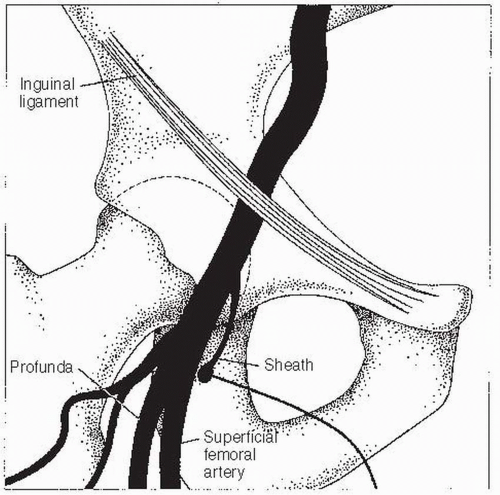
FIGURE 64.1 Landmarks for right femoral artery puncture.


B. Brachial and radial approach.

In certain patients, it may be desirable to perform the catheterization by a brachial or radial route, for which specialized equipment is available. Percutaneous brachial or radial access is similar to the femoral approach described above. In addition, a surgical cut-down was historically performed to enter the brachial arteries under direct visualization, though surgical cut-downs are rarely performed in the modern era. For a left brachial approach, Judkins catheters are adequate. For a right brachial approach, Amplatz or multipurpose catheters are used, although it may be difficult to engage a left internal mammary artery (LIMA) graft from the right arm; a specially designed brachial internal mammary artery catheter is available for the latter purpose. In rare circumstances, an axillary approach can be used, though the rate of neurovascular complications is higher.

Increasingly, operators now use the radial approach as the default approach. The radial approach has been associated with fewer bleeding complications when compared with the femoral approach and does not require a long period of immobilization of the patient afterward. It is thus preferred by patients. Radiation exposure data have been conflicting and can be influenced by multiple factors including the familiarity of the operator with the radial approach. To obtain vascular access from the radial site, the Allen test should be performed prior to radial artery catheterization. The patient’s arm is abducted at a 70° angle and the wrist is hyperextended. The site is prepped and draped in a manner similar to the femoral and brachial access sites. Local anesthetic is injected. The radial site should be roughly 1 cm above the styloid process. Either an 18G needle or a micropuncture needle (22G) is inserted at 30° to 45° into the radial artery. A sheath is advanced in the same manner as described above using the Seldinger technique. Local infusions of nitroglycerin and/or verapamil can be injected to decrease radial artery spasm. Heparin 3,000 to 5,000 IU should be considered to avoid sheath thrombosis. Once access is obtained, a similar process of advancing a catheter over a guidewire is performed as in other access sites. Diagnostic and interventional procedures can be performed via the radial artery using traditional catheters as well as a number of newer catheters designed specifically for radial access such as the Jacky catheter or the Tiger catheter.


C. Special situations.

In patients with prosthetic femoral grafts, it is preferable to use a dilator first before placing the sheath to prevent the sheath from kinking as it passes through the graft. This technique is also useful in obese patients. If a synthetic graft is old, fluoroscopy can be performed to determine if the graft is heavily calcified—a sign that it may not seal well after sheath removal. In patients with tortuous or diseased vessels, a Wholey wire or Terumo glidewire can be used to get catheters up the aorta. If marked iliac tortuosity is present and causes inability to torque catheters, a long sheath can be used to straighten out the iliac vessel. At times, a stiffer wire (such as an Amplatz wire) can provide better support to advance catheters. In patients with an AAA for whom a femoral approach is chosen, exchange wires should be used for every change of catheter. Use of a softer wire (such as a Wholey wire) can prove less traumatic to the vessel, as can use of a JR 4 to direct the guidewire.


D. Catheters.

The catheters commonly used for coronary angiography include the Judkins and the Amplatz systems. For the left coronary artery (LCA), the size of the Judkins left (JL) catheters ranges from JL 3.5 to JL 6. The Amplatz left (AL) catheters used commonly range in size from AL I to AL III (Fig. 64.2 shows the shapes of the JL and AL catheters). Similarly for the right coronary artery (RCA), the Judkins right (JR) catheters range in size from JR 3.5 to JR 6. The Amplatz right (AR) catheters commonly used range from AR I to AR III. In addition, there is also an AR-modified catheter (Fig. 64.3 shows the shapes of the JR and AR coronary catheters commonly used). Other catheters used include the multipurpose catheters (multipurpose A1, A2, B1, and B2 catheters), which can be used for cannulating the left and right coronary arteries and bypass grafts. For coronary bypass grafts, the right or the left coronary bypass catheters may be used. The internal mammary artery may be cannulated using either the internal mammary or the internal mammary special catheters (see Fig. 64.4).


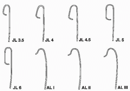
Figure 64.2 Catheters used for cannulating the left coronary artery. JL, Judkins left; AL, Amplatz left.


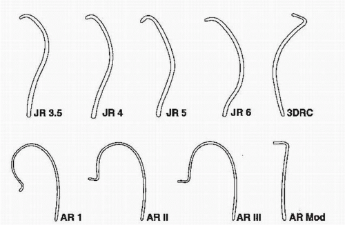
FIGURE 64.3 Catheters used for cannulating the right coronary artery. JR, Judkins right; AR, Amplatz right; 3DRC, no torque right coronary catheter; Mod, modified.


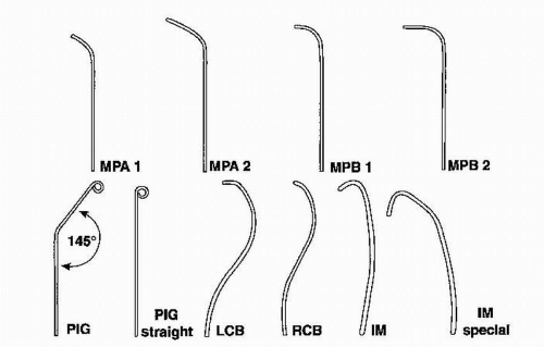
FIGURE 64.4 Other catheters used for cardiac catheterization. MP, multipurpose; PIG, pigtail; LCB, left coronary bypass catheter; RCB, right coronary bypass catheter; IM, internal mammary.


Jun 7, 2016 | Posted by in CARDIOLOGY | Comments Off on Left Heart Catheterization

Full access? Get Clinical Tree

Get Clinical Tree app for offline access