Left Atrial Enlargement
Gregory Kicska, MD, PhD
DIFFERENTIAL DIAGNOSIS
Common
Left Heart Failure
Mitral Valve Disease
Chronic Atrial Fibrillation
Less Common
Left to Right Shunts
Rare but Important
Constrictive Pericarditis/Restrictive Cardiomyopathy
ESSENTIAL INFORMATION
Key Differential Diagnosis Issues
Radiograph: Double density sign, splaying of carina, superior displacement of left main bronchus, posterior esophageal displacement, enlarged LA appendage
Aortic root diameter: LA short axis ratio should be near 1:1
Rightward displacement of interatrial septum suggests LA enlargement
Normal volume = 22 ± 5 mL/m2
Helpful Clues for Common Diagnoses
Left Heart Failure
Chronic ischemia, diabetes, and chronic hypertension most common etiologies
Diastolic heart failure can exist with normal LV end diastolic volume and ejection fraction
Mitral Valve Disease
Stenosis
Coexistent edema suggests valve area is less than 1 cm2 (normal 4-6 cm2)
Calcified leaflets not to be confused with mitral annular calcification
Regurgitation
Often coexists with stenosis and calcified valve
Absence of calcifications suggests prolapse or ruptured papillary muscle
Chronic Atrial Fibrillation
Exclude LA appendage thrombus on contrast exams
Senescent dilation may lead to A-fib
Helpful Clues for Less Common Diagnoses
Left to Right Shunts
Qp:Qs ratio does not equal 1
VSD does not cause LA dilation unless large
ASD only with Eisenmenger physiology in advanced age
PDA will also have LV enlargement
Helpful Clues for Rare Diagnoses
Constrictive Pericarditis/Restrictive Cardiomyopathy
Tubular-shaped ventricles are disproportionally smaller than atria
Constrictive pericarditis suggested by focal or diffuse pericardial thickening > 4 mm or calcification in presence of heart failure
Restrictive cardiomyopathy suspected in absence of pericardial thickening
Image Gallery
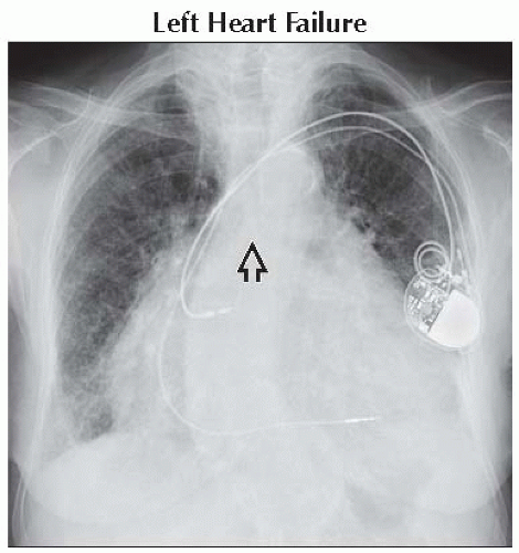 Frontal radiograph shows cardiomegaly with left atrial and ventricular enlargement in a patient with heart failure. Note splaying of the carinal angle (normal < 90°)
 . .Stay updated, free articles. Join our Telegram channel
Full access? Get Clinical Tree
 Get Clinical Tree app for offline access

|