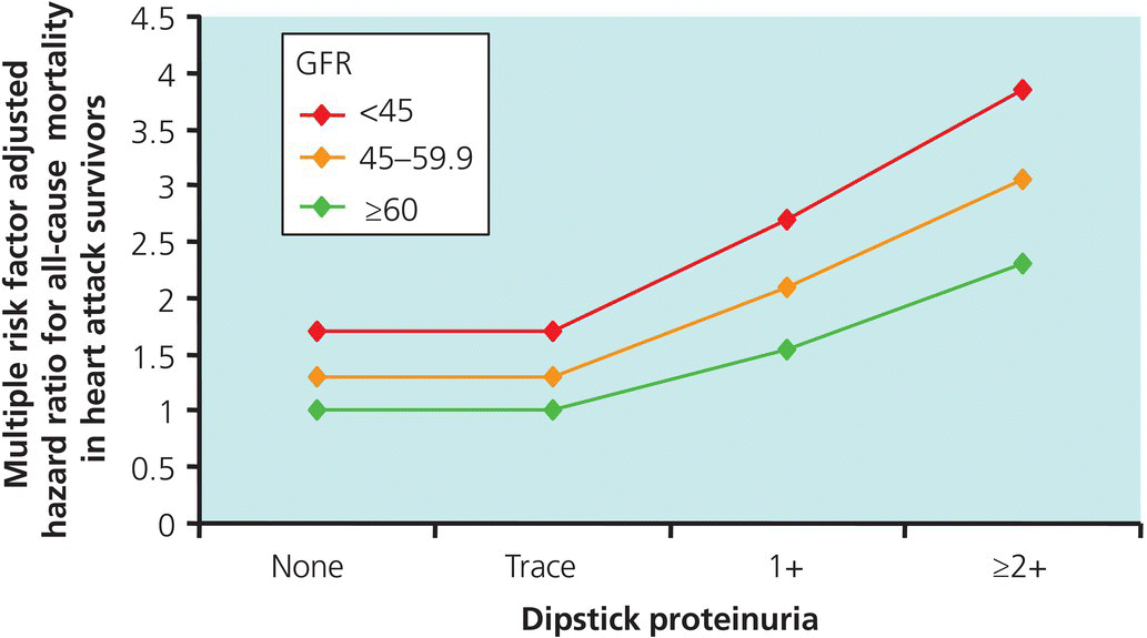
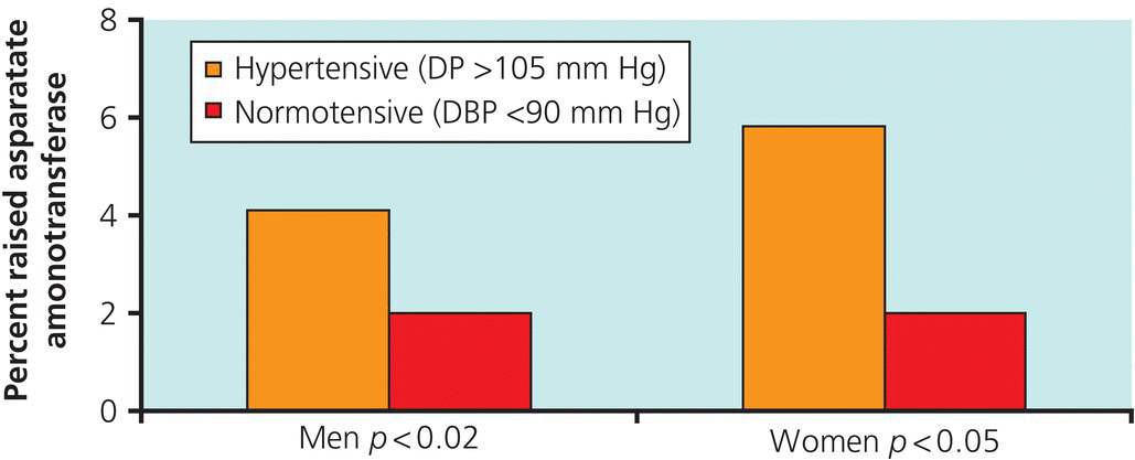
Chapter 7 Investigations are needed in all patients with hypertension to detect any underlying cause (e.g. to exclude secondary hypertension), assess for the consequences of hypertension (target organ damage) and test for other cardiovascular risk factors (Table 7.1). Very thorough investigations should be performed in young patients (those younger than 40 years), patients who present with severe or resistant hypertension and patients in whom secondary hypertension is suspected. Chestxray, urine microscopy and culture and echocardiography are not ‘routine’ investigations and are not needed in most patients. The NICE guidelines suggest the following routine tests. Table 7.1 BHS/NICE guidance for the initial assessment for target organ damage in hypertensive patients Some of these tests can be conducted in primary care. In addition to the BHS/NICE recommendations above, we strongly advocate the routine dipstick testing of the urine in all hypertensive patients in primary and secondary care. Proteinuria and microscopic haematuria suggest intrinsic renal disease, including glomerulonephritis, particularly immunoglobulin A (IgA) nephropathy, autosomal dominant polycystic kidney disease (ADPKD) or pyelonephritis. In patients with proteinuria, the risk of mortality and morbidity is roughly doubled for a given blood pressure. Proteinuria is a powerful predictor of all-cause mortality in hypertensive patients with or without cardiovascular damage. This effect is independent of the amount of renal impairment (Figure 7.1). Figure 7.1 The relationship between dipstick proteinuria and all-cause mortality in patients with existing coronary heart disease. Similar trends are found in patients with no end-organ damage. Source: Data from Tonelli, M, et al. (2006) British Medical Journal, 332, 1426–1429. Microalbuminuria (urine albumin less than 300 mg/24 h) or urine albumin: creatinine ratio also predicts all-cause and cardiovascular mortality. By contrast, microscopic haematuria does not appear to predict end-organ damage. It may occur in severe uncomplicated essential hypertension. However, it may be a marker for underlying primary renal disease. When haematuria is persistent and marked, renal and renal tract malignancies should be excluded and urological referral may be necessary. Serum levels of potassium are usually low or low-normal in patients with primary hyperaldosteronism (Conn’s syndrome). This is often associated with high or high-normal serum levels of sodium. Low levels of potassium sometimes occur in patients who take diuretics, but usually, thiazide diuretics only lower potassium by 0.5 mmol/l. If hypokalaemia is marked, it is possible that it is related to underlying aldosterone excess. Patients with malignant hypertension may have mild hypokalaemia because of aldosterone excess, secondary to high levels of renin caused by juxtaglomerular cell ischaemia. As blood pressure is brought under control, this hypokalaemia often normalises. If serum levels of potassium remain low without diuretic treatment despite good control of blood pressure over a few months, primary hyperaldosteronism should be excluded. Hyperkalaemia may develop in patients with renal failure and those who take drugs such as angiotensin-converting enzyme inhibitors, angiotensin receptor blockers and potassium sparing diuretics (e.g. spironolactone and amiloride). Spironolactone is increasingly used as third-line treatment in patients with resistant hypertension, and potassium and renal function need to be monitored regularly. Low table salt alternatives, which contain potassium salts, should be used cautiously with angiotensin-blocking drugs as dangerous hyperkalaemia can occur, particularly if spironolactone is also being used. This is a problem which is becoming more common as angiotensin-blocking drugs and spironolactone are frequently used together in both hypertension and heart failure. Serum levels of sodium may be high or high-normal in patients with primary hyperaldosteronism (Conn’s syndrome). This is usually associated with low or low-normal serum levels of potassium. In patients with secondary hyperaldosteronism as a result of malignant hypertension or renal disease, serum levels of sodium can be low or low-normal. Low levels of sodium may also be present with overuse of diuretics. Profound hyponatraemia, which may cause confusion and hypotension, is occasionally seen in patients even on low doses of diuretics. Non-malignant essential hypertension only rarely causes renal impairment, but associated co-morbid disease (such as diabetes mellitus) and concomitant treatment with angiotensin-converting enzyme inhibitors or angiotensin receptor blockers can lead to renal impairment. Almost all intrinsic renal diseases can cause hypertension, and levels of urea and creatinine in serum should be part of the initial work up in a patient with newly diagnosed hypertension. Even a modest increase in levels of creatinine in serum needs more detailed investigation. In recent years, it has become fashionable to assess renal function using a formula devised from the US Modification of Diet in Renal Disease (MDRD) study. This has proved a little controversial. It has long been recognised that serum creatinine levels do not rise above the so-called normal range until renal function is about halved. The MDRD study attempted to correct for this. The estimated glomerular filtration rate (eGFR) formula takes into account age, gender, body weight (as a surrogate for muscle bulk) and ethnicity (African American vs. non-African American). The grade of renal impairment can then be calculated but this should also take into account the presence of other abnormalities including proteinuria and haematuria. Taking these criteria, patients with a eGFR above 90 ml/min (excellent) will be labeled as having grade 1 chronic kidney disease (CKD) (Table 7.2). It would appear that all patients, regardless of their eGFR, have some form of CKD. Table 7.2 The MDRD classification of chronic kidney disease This has presented a particular problem in older patients with serum creatinine levels that are about average or near average for their age. Once labeled as having CKD, they are subjected to renal imaging and often unnecessary nephrological referral. It is to be hoped that the use of serum creatinine levels to measure eGFR will be refined over the coming years. High levels of uric acid in serum are found in about 40% of patients with hypertension, especially in association with renal impairment. Increased ingestion of alcohol and the use of thiazide diuretics can also lead to higher levels of uric acid. Whether high levels of uric acid are harmful in their own right, or only by association with hypertension, hyperlipidaemia and diabetes remains uncertain. There is no information as to whether the treatment of hyperuricaemia with allopurinol in symptomless hypertensive patients with normal renal function is of any value. There is an increased prevalence of abnormal liver function tests (LFTs) in patients with hypertension. (Figure 7.2). This is partly explained by the association between excess alcohol intake in the causation of hypertension. However, abnormal LFT are also seen in people who consume no alcohol at all. Such patients are often overweight and/or have type 2 diabetes, as part of Non-Alcoholic Fatty Liver Disease (NAFLD). This syndrome is usually reversible with weight reduction but may progress to Non-Alcoholic Steato-Hepatitis (NASH) or even cirrhosis. Figure 7.2 Serum aspartate aminotransferase in normotensives and hypertensives in the Renfrew Community study. Source: Data from Beevers, D.G. (1977) Lancet, 2, 114–115.
Investigation in patients with hypertension
Routine investigations
Test for the presence of protein in the urine by sending aurine sample for estimation of the albumin: creatinine ratio and test for haematuria using a reagent strip.
Take a blood sample to measure plasma glucose, electrolytes, creatinine, estimated glomerular filtration rate (eGFR), serum total cholesterol and HDL cholesterol.
Examine the fundi for the presence of hypertensive retinopathy.
Arrange for a12-lead electrocardiograph to be performed.
Urinalysis

Biochemistry
Serum potassium
Serum sodium
Serum urea and creatinine
Estimated glomerular filtration rate
eGFR
CKD stage
>90 ml/min
Without other abnormality otherwise regard as normal
1
60–89 ml/min
With other abnormality otherwise regard as normal
2
30–59 ml/min
Moderate impairment
3
15–29 ml/min
Severe impairment
4
<15 ml/min
Established renal failure
5
Serum uric acid
Liver function tests

Investigation in patients with hypertension
Source: Reproduced with permission from Krause, T., et al. (2011) British Medical Journal, 343, d4891. © BMJ Publishing Group Ltd.
Only gold members can continue reading. Log In or Register to continue

Full access? Get Clinical Tree


