Fig. 1.1
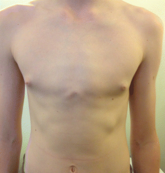
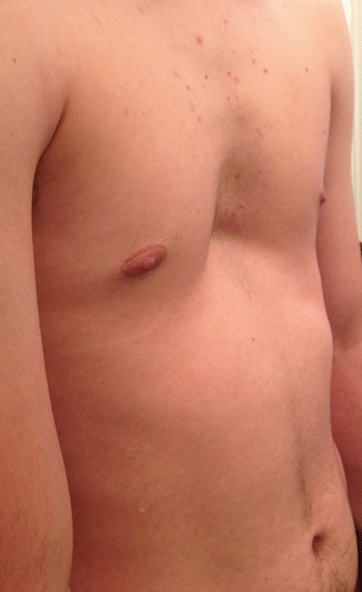
Pectus excavatum
The compression of the sternum limits thoracic volume and therefore vital capacity, negatively impacting exercise tolerance and endurance during cardiovascular exercise. In some cases, cardiac compression is observed. This causes a significant reduction in cardiac output further contributing to exercise intolerance and fatigue.
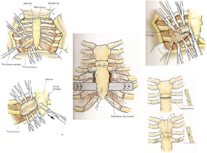
Surgical intervention has significantly benefitted patient respiratory function and exercise tolerance. Initially, the deformity was surgically corrected through the Ravitch procedure. Now, more commonly, the Nuss procedure is undertaken to readjust and advance the sternal position with the use of a concave steel bar inserted retrosternal through bilateral incisions. The intervention has very few documented side effects but causes marginal postoperative pain that varies amongst individuals. The pain is usually mild and short-lasting, however, effective pain management greatly influences a patient’s satisfaction and perspective on the success of the treatment. Pain management differs amongst institutions with the majority using thoracic epidurals. Few institutions utilise patient controlled anaesthesia and these centres believe that it should become the more widely used option postoperatively as it decreases the length of hospitalisation after the intervention (Fig. 1.2).


Fig. 1.2
Ravitch procedure
The introduction of the NUSS procedure in 1998 for the surgical correction of pectus excavatum was the beginning of a new era for the management of chest wall deformities and a new significant chapter in the modern Thoracic Surgery [1]. The ‘minimally-invasive’ Nuss procedure is growing in popularity due to infrequent complications, very few side effects and a short length of hospitalisation, lasting 2–4 days, post-operation (Fig. 1.3).


Fig. 1.3
NUSS procedure
Furthermore, pectus excavatum has profound effects on the psychological state of the individual suffering with the deformity. Pectus excavatum patients suffer frequent embarrassment over physical appearance and teasing by childhood peers. The typical patient starts to become aware of the condition at the onset of puberty and this has detrimental effects on the individual’s confidence and happiness in early adolescence. In fact, 80 % of patients undergoing treatment admitted to suffering with psychological limitations concerning attractiveness, self-esteem and somatization. In severe cases, some individuals may retreat from society and cease to socialize with peers or participate in exposing sporting activities, such as swimming. This led to the labeling of pectus excavatum as a psychosomatic disorder and further merited surgical and non-surgical intervention.
Over the decades different studies revealed that most of deformities are familiar with a strong genetic involvement and usually related with other syndromes, anomalies and defects making the management challenging [2]. Nevertheless the majority of chest wall anomalies remain rare clinical entities and some of them like thoracic ectopia cordis are not compatible with life and very unlikely to be benefited by a surgical procedure [3]. The approach of chest wall deformities is still controversial as it’s not the clinical symptoms – mainly cardiopulmonary – but also the psychosocial aspects and effects of poor cosmetic that have a huge impact to the quality of life [4]. For that reason in recent years has been a significant increase in the interest of clinicians for assessment and management of these patients. Toward that direction new assessment criteria have been established and new minimally invasive surgical techniques have been introduced. Different classifications have been proposed through years to categories chest wall anomalies. In 2006 Acastello classified them into five types depending on the site of origin of the anomaly (type I: cartilagineous, type II: costal, type III: chondro-costal, type IV: sternal, type V: clavicle- scapular) [5].
Among all chest wall deformities Pectus Excavatum (PE) or funnel chest represents the most common congenital chest wall deformity accounting for 90 % of all deformities [6]. The first description came from Bauhinus1 in the sixteenth century [7] and main characteristic is the depression of sternum and lower cartilages [8] with an incidence between 1 and 8 per 1000 children [9].
Pectus Carinatum (PC) or protrusion deformity of the chest wall accounts for 5 % of all chest wall deformities affecting 1 in 2500 live births [10]. It can be unilateral, bilateral or mixed and there is predominance in males (Fig. 1.4) [11].


Fig. 1.4
Pectus carinatum
Pectus arcuatum represents a rare category of chest wall deformities in the family of pectus anomalies and It includes mixed excavatum and carinatum features along a longitudinal or transversal axis resulting in a multiplanar curvature of the sternum and adjacent ribs (Fig. 1.5) [12].


