Fig. 6.1
Haller Index on CT scan
Chest X-ray
Evaluation of patients with plain radiographs is performed in two projections, frontal and lateral [15, 16]. PE deformity can produce characteristic findings of right middle lobe opacification on the frontal view that can often be mistaken for right middle lobe pneumonia or atelectasis [15]. This abnormal appearance is in fact due to compression of anterior chest wall soft tissue [15]. Other features that may also be apparent on frontal projections include displacement of the cardiac silhouette to the left in more than 50 % of patients [17]. Takahashi et al. found obliteration of the descending aortic interface in 30 % of PE cases reviewed retrospectively in a cohort of 70 patients. Although no significant relationship was found between this and the severity of thoracic deformity, an indirect correlation to cardiac rotation angle does appear evident [18].
Lateral projections demonstrate posterior displacement of the sternum that is evident as an opacity filling the retrosternal space with ribs projecting anterior to the sternum, thus confirming PE deformity (Figs. 6.2, 6.3, 6.4, and 6.5) [15].
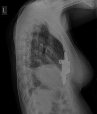
Fig. 6.2
Chest x-ray PA view
Fig. 6.3
Chest x-ray with left lateral view
Fig. 6.4
Chest x-ray left lateral with the bar
Fig. 6.5
Chest x-ray with the bar
CT Scan
Computerised tomography (CT) scans have long been the primary imaging modality for evaluation of PE deformity. As described earlier, the Haller Index for objectively grading the severity of PE deformity was developed through the use of CT scans by dividing the transverse diameter of the chest by the anteroposterior diameter at the point of maximal sternal depression. In addition, CT scans can effectively demonstrate the degree of cardiac compression and displacement and its relationship to the sternum, the degree of pulmonary compression and atelectasis, asymmetry of the chest, sternal torsion and compensatory development of a barrel chest (Figs. 6.6, 6.7, 6.8, 6.9, 6.10, and 6.11) [2, 19].
Fig. 6.6
CT scan with minimal depression and displacement
Fig. 6.7
CT scan with tilt of sternum with depressed right side
Fig. 6.8
CT scan with depression more on left side
Fig. 6.9
CT scan with leftward tilt and depression
Fig. 6.10
3D reconstruction from CT showing the sternal depression and ribs alignment (a) lateral view; (b) oblique view
Fig. 6.11
3D reconstruction from CT showing the sternal depression and ribs alignment oblique view (a) right oblique view; (b) left oblique view
MRI and CMR
Certain centres have adopted Magnetic Resonance Imaging as the primary imaging modality to calculate the Haller Index. The primary advantage over CT is the absence of ionising radiation [10]. Piccolo et al. demonstrated strong comparability of Haller Indices and Asymmetry Indices obtained using fast MRI and CT scanning. In addition, fast MRI demonstrates excellent soft tissue contrast and thus high quality assessment of cardiac displacement or rotation, great vessel anomalies and asymmetric volume between left and right hemithorax (Fig. 6.12) [20, 21].
Fig. 6.12
MRI scan transverse section showing sternal depression
CMR has also been investigated as a potential imaging modality with the aim to delineate the anatomical and physiological components of pectus excavatum in addition to calculating the Haller Index. Saleh et al. demonstrated statistically significant higher HI in PE patients compared to non pectus patients (9.6 versus 2.8). The group were also able to demonstrate significant left lateral shift of the heart compared to controls (84 % versus 64 %) and reduction in right ventricular ejection fraction, which may be suggestive of changes in myocardial performance [22]. Humphries et al. have also demonstrated similar results with calculated HI of PE and non pectus patients [23].
Echocardiography
Echocardiograms are performed to search for the presence of major valvular pathology and chamber compression (Fig. 6.13). The right ventricle and atrium can be compressed by the sternum resulting in problems during diastolic filling [20].
Fig. 6.13
Echocardiogram with four-channel view
Summary
Recent discussion has questioned the absolute necessity of performing a CT scan for evaluation of PE deformity. The main factor in searching for alternative imaging techniques is the high doses of radiation that are being delivered by CT scans to patients in their adolescence or younger who are being assessed for PE deformity. Four independent groups have evaluated the use of two-view chest x-rays in the pre-operative assessment of patients with PE deformity and conclude strong comparability of HI calculated from chest x-rays with that of CT scans and strong interobserver correlation. The unanimous recommendations from these studies is to replace CT imaging with chest X rays as the primary imaging modality for PE surgical workup, which would result in a 100 fold reduction in effective radiation dose [1, 24–26]. The counter argument to this is that chest x-rays provide no information regarding sternal asymmetry or torsion or the degree of cardiac compression and displacement [19]. One study concluded that no additional information was gleaned from a CT scan that was not already evident from two-view CXRs and instead adopted the use of low dose CT scanning comprising of five to seven slices through the point of maximal sternal depression in addition to two view CXRs. This has reduced radiation doses by up to 80 % [27] while still providing comprehensive visualisation of the local anatomy. In addition to significantly reducing the radiation exposure a substantial cost saving is also achievable when employing this strategy for pre-operative assessment [24].
Other groups have investigated the use of MRI and CMR scanning as the primary imaging modality and thus eliminating ionising radiation exposure completely. Two separate studies have shown favourable results for using MRI over CT scans to calculate the HI and AI while detailing anatomical information such as displacement, rotation or compression of the heart or great vessel anomalies [10, 21]. In addition to no radiation exposure the images can be acquired in less than 5 min without any compromise in quality. Further research demonstrates that cine MR can provide additional diagnostic information through evaluation of chest morphology and chest wall kinetics [10]. CMR has also been shown to be beneficial for the same reasons as MRI but also in its ability to provide accurate and dynamic assessment of cardiovascular function and thus potentially eliminating the need for echocardiographic studies.
It is clear that a growing number of institutions are moving away from the use of full chest CT scanning as the primary modality for calculating HI and AI in patients with PE deformity and adopting imaging strategies that have lower or no radiation exposure. All of the studies performed with alternative imaging modalities to CT have a smaller cohort of patients but so far the results are encouraging.
Pectus Carinatum
Pectus carinatum (PC) is the second most common anterior chest wall anomaly occurring in approximately one in 1500 live births with a male to female ratio of 4:1 [20, 28]. It is characterised by sternal and costal cartilage elevation [20] and depending on the area involved can be classified into two variant types [20, 28].
Chondrogladiolar subtype is the more common deformity involving protrusion of the mid to lower portion of the anterior chest wall and inferior costal cartliages and part of the gladiolus [28, 29]. Chondromanubrial deformity or Currarino-Silverman syndrome in cases with associated congenital heart disease, is the less common subtype involving protrusion of the second and third costal cartilages with elevation of the sterno-manubrial joint [20, 28].
Pre-treatment radiological evaluation of PC aims to establish the presence of an increased antero-posterior (AP) diameter in association with protrusion of the sternum [28]. Features that may be apparent on plain chest radiography include a narrowed thorax and scoliosis on posteroanterior projections. Lateral projections provide a more striking image of the deformity. Features that can be identified include the presence of a bowed, short, comma-shaped sternum with an increased AP diameter [28]. Further differentiation of PC into its variant forms, as determined by the site of sternal protrusion, can also be adequately demonstrated on lateral projections as shown by various groups [28–31]. Computed tomography (CT) has also been advocated for use in the evaluation of patients with PC as part of the routine pre-treatment assessment (Fig. 6.14) [20]. Desmaris et al. conclude that CT scanning should be reserved for cases with ‘mixed’ deformities, where sternal angle measurements can be easily calculated [30]. More recently Lee et al. presented their experience with the use of MRI for evaluation of PC. Early results show MRI to be highly effective at measuring sternal angle rotation and asymmetry index in PC patients, which can be used in pre-treatment evaluations [32].