The purpose of the present study was to assess the impact of clinical presentation and pretest likelihood on the relation between coronary calcium score (CCS) and computed tomographic coronary angiography (CTA) to determine the role of CCS as a gatekeeper to CTA in patients presenting with chest pain. In 576 patients with suspected coronary artery disease (CAD), CCS and CTA were performed. CCS was categorized as 0, 1 to 400, and >400. On CT angiogram the presence of significant CAD (≥50% luminal narrowing) was determined. Significant CAD was observed in 14 of 242 patients (5.8%) with CCS 0, in 94 of 260 patients (36.2%) with CCS 1 to 400, and in 60 of 74 patients (81.1%) with CCS >400. In patients with CCS 0, prevalence of significant CAD increased from 3.9% to 4.1% and 14.3% in nonanginal, atypical, and typical chest pain, respectively, and from 3.4% to 3.9% and 27.3% with a low, intermediate, and high pretest likelihood, respectively. In patients with CCS 1 to 400, prevalence of significant CAD increased from 27.4% to 34.7% and 51.7% in nonanginal, atypical, and typical chest pain, respectively, and from 15.4% to 35.6% and 50% in low, intermediate, and high pretest likelihood, respectively. In patients with CCS >400, prevalence of significant CAD on CT angiogram remained high (>72%) regardless of clinical presentation and pretest likelihood. In conclusion, the relation between CCS and CTA is influenced by clinical presentation and pretest likelihood. These factors should be taken into account when using CCS as a gatekeeper for CTA.
Noncontrast-enhanced computed tomography visualizes coronary calcium as a marker for coronary artery disease (CAD) and quantifies the presence and extent of coronary calcium by use of the coronary calcium score (CCS). More recently, contrast-enhanced computed tomographic coronary angiography (CTA) has been introduced. This technique provides direct visualization of the coronary arteries and allows more detailed assessment of coronary atherosclerosis and stenosis severity. Several studies have suggested that CCS might be useful as a gatekeeper to CTA in diagnosis of significant CAD in patients presenting with chest pain. Absence of calcium could exclude the presence of significant CAD, indicating no need for further imaging, whereas patients with an increased CCS could be referred for CTA for additional information on stenosis severity. To evaluate the feasibility of such an approach, several comparative studies have been performed addressing the relation between CCS and CTA in patients presenting with chest pain. However, large discrepancies have been observed, which have been ascribed to differences in clinical characteristics of the studied populations. The purpose of the present study therefore was to systematically assess the impact of clinical presentation and pretest likelihood on the relation between CCS and CTA to determine the role of CCS as a gatekeeper to CTA for diagnosis of significant CAD in patients presenting with chest pain.
Methods
The study population consisted of patients with suspected CAD who were clinically referred for further cardiac assessment because of chest pain. The included patients underwent CCS scanning and CTA. Exclusion criteria were cardiac arrhythmias, renal insufficiency (defined as glomerular filtration rate <30 ml/min), known hypersensitivity to iodine contrast media, severe claustrophobia, and pregnancy. In addition, patients with an uninterpretable CT angiographic examination were excluded. Symptoms were classified as typical angina, atypical angina, or nonanginal chest pain. Typical anginal chest pain was defined as combination of (1) discomfort in the anterior chest, neck, shoulders, jaw, or arms; (2) precipitated by physical exertion or emotional stress; and (3) relieved by rest or nitroglycerin within minutes. Atypical chest pain was defined as chest pain with 2 of these 3 factors and nonanginal chest pain was defined as chest pain with <2 of these 3 factors. Pretest likelihood was defined according to criteria of Diamond and Forrester, which are based on previously observed prevalences of significant CAD in age, gender, and chest pain subgroups. Thresholds for low, intermediate, and high pretest likelihood were ≤13.4, 13.5 to 87.2, and ≥87.3, respectively.
The examination was performed using a 64-detector row helical scanner (Aquilion 64; Toshiba Multislice System, Toshiba Medical Systems, Otawara, Japan) or a 320-detector row volumetric scanner (Aquilion ONE, Toshiba Medical Systems). Before CCS and CT angiographic examinations, patients’ heart rate and blood pressure were monitored. In the absence of contraindications, patients with a heart rate exceeding the threshold of 65 beats/min were administered β-blocking medication (metoprolol 50 to 100 mg orally). Before the helical scan, a nonenhanced low-dose electrocardiographically gated scan was performed to measure CCS. The CCS scan was prospectively triggered at 70% or 75% of RR interval and performed using the scan parameters 4- × 3.0-mm or 2.5-mm collimation for 64-row computed tomography and single-rotation-wide volume acquisition (320 × 0.5 mm, reconstructed to 3-mm slices) for 320-row computed tomography with a gantry rotation time 350 to 500 ms, tube voltage 120 kV, and tube current 200 to 250 mA.
For the 64-row contrast-enhanced scan, collimation was 64 × 0.5 mm, tube voltage 100 to 135 kV, and tube current 250 to 350 mA, depending on body mass index and thoracic geometry. Nonionic contrast material (Iomeron 400; Bracco, Milan, Italy) was administered with an amount of 80 to 110 ml followed by a saline flush at a flow rate of 5 ml/s. For the 320-row contrast-enhanced scan the heart was imaged in a single heartbeat using prospective triggering with exposure interval depending on heart rate. Scan parameters were 320- × 0.5-mm collimation, 350-ms gantry rotation time, 100- to 135-kV tube voltage, and 400- to 580-mA tube current, depending on body mass index. In total, contrast material 60 to 90 ml was administered at a rate of 5 to 6 ml/s followed by a saline flush.
Postprocessing of CCS and CT angiographic examinations was performed on dedicated workstations (Vitrea 2.0 or Vitrea FX 1.0, Vital Images, Minnetonka, Minnesota). CCS was calculated using the Agatston method and patients were categorized as CCS 0, CCS 1 to 400, and CCS >400. CT angiograms were examined using axial slices, curved multiplanar reconstructions, and maximum intensity projections. All CT angiographic scans were interpreted by 2 experienced observers blinded to the results of CCS. CT angiographic examinations were classified according to the most severe lesion. In each patient, presence of CAD was determined. Further differentiation was made between nonsignificant and significant CAD using a diameter stenosis ≥50% as a threshold for significant lesions.
Continuous variables were expressed as mean ± SD and categorical baseline data were expressed in numbers and percentages. Differences in baseline clinical variables among the CCS subgroups were compared using analysis of variance and Student’s t and chi-square tests. Prevalence of significant CAD on CT angiogram in each CCS category was determined according clinical presentation and pretest likelihood. All statistical analyses were performed using SPSS 16.0 (SPSS, Inc., Chicago, Illinois).
Results
The study population consisted of 602 patients presenting with chest pain who had undergone CCS and CTA. In 26 of these patients (4.3%), CT angiographic examination was uninterpretable because of the presence of motion artifacts, increased noise owing to a high body mass index, and breathing. After exclusion of these patients, 576 remained for further analysis. Baseline characteristics of the patient population are presented in Table 1 .
| Variable | All (n = 576) | CCS | p Value | ||
|---|---|---|---|---|---|
| 0 | 1–400 | >400 | |||
| (n = 242) | (n = 260) | (n = 74) | |||
| Men | 273 (47%) | 93 (38%) | 137 (53%) | 43 (58%) | 0.001 |
| Age (years) | 56 ± 12 | 50 ± 11 | 59 ± 11 | 66 ± 9 | <0.001 |
| Diabetes mellitus | 105 (18%) | 33 (14%) | 49 (19%) | 23 (31%) | 0.003 |
| Hypertension | 254 (44%) | 66 (27%) | 136 (52%) | 52 (70%) | <0.001 |
| Hypercholesterolemia ⁎ | 199 (35%) | 57 (24%) | 106 (41%) | 36 (49%) | <0.001 |
| Current smokers | 115 (20%) | 49 (20%) | 40 (15%) | 26 (35%) | 0.001 |
| Body mass index ≥30 kg/m 2 | 109 (19%) | 43 (18%) | 48 (19%) | 18 (24%) | 0.48 |
| Symptoms | 0.017 | ||||
| Nonanginal chest pain | 205 (36%) | 103 (43%) | 84 (32%) | 18 (24%) | 0.005 |
| Atypical chest pain | 249 (43%) | 97 (40%) | 118 (45%) | 34 (46%) | 0.42 |
| Typical chest pain | 122 (21%) | 42 (17%) | 58 (22%) | 22 (30%) | 0.06 |
| Pretest likelihood † | <0.001 | ||||
| Low | 117 (20%) | 89 (37%) | 26 (10%) | 2 (3%) | <0.001 |
| Intermediate | 370 (64%) | 131 (54%) | 188 (72%) | 51 (69%) | <0.001 |
| High | 89 (16%) | 22 (9%) | 46 (18%) | 21 (28%) | <0.001 |
⁎ Serum total cholesterol ≥230 mg/dl and/or serum triglycerides ≥200 mg/dl or use of lipid-lowering agents.
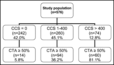
Median CCS of the study population was 7 (25th to 75th percentiles 0 to 133). Calcium was absent in 242 patients (42%), a CCS of 1 to 400 was present in 260 patients (45.1%), and a CCS >400 in 74 patients (12.8%). Significant CAD was observed on CT angiogram in 168 patients (29%). In the remaining 408 patients (71%), nonsignificant CAD was observed in 184 patients (32%) and 224 patients (39%) were classified as normal.
Figure 1 illustrates the CT angiographic findings in the different CCS groups. In patients without any coronary calcium (CCS 0), significant CAD was observed in 14 patients (5.8%). In the group of patients with a CCS of 1 to 400, 94 patients (36.2%) had significant CAD on CT angiogram. In patients with a high CCS >400, significant CAD was observed in 60 patients (81.1%).
Figures 2 and 3 illustrate the prevalence of significant CAD in the different CCS groups according to clinical presentation and pretest likelihood.


