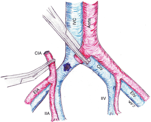
Fig. 20.1
Normal anatomy of the iliac arteries and veins. IVC inferior vena cava, CIV common iliac vein, EIV external iliac vein, IIV internal iliac vein, CIA common iliac artery, EIA external iliac artery, IIA internal iliac artery
Given the anatomically protected location of the iliac vessels, injuries with enough force to cause iliac vessel injuries are frequently complicated by associated damage to surrounding organs [1]. Many patients will have more than one vascular injury and may have combined arterial and venous injuries. In patients with penetrating trauma to the iliac vein, 34–57 % will have a concomitant injury to the iliac artery [4–6, 10]. Associated nonvascular injuries occur in almost all patients and most frequently include the small bowel, colon, bladder, and ureter (Table 20.1).
Table 20.1
Associated injuries with iliac vein injury
Small bowel | 46.4–77.6 % |
Colon | 24.0–57.1 % |
Bladder | 6.1–10.2 % |
Ureter | 3.9–10.1 % |
Liver | 1.7–12.0 % |
Kidney | 1.1–4.1 % |
Pelvic fracture | 1.1–6.7 % |
20.4 Clinical Assessment
On initial evaluation, patients are often hypotensive and tachycardic secondary to blood loss following either penetrating or blunt trauma. Full exposure is required to identify penetrating injuries. With blunt trauma, the diagnosis is especially challenging but is often associated with pelvic fractures. The most critical component to patient survival is rapid recognition of hemorrhagic shock and rapid transport to a trauma center with notification of the trauma surgery team.
20.5 Diagnostic Testing
Laboratory testing frequently demonstrates mild acidosis and anemia [5]. If the diagnosis of iliac vessel injury is delayed, the patient will become progressively more acidotic, anemic, and coagulopathic. Iliac vein injuries can present with retroperitoneal hematoma, free intraperitoneal hemorrhage, or both. The Focused Assessment with Sonography for Trauma (FAST) exam is commonly employed in most major trauma centers to detect major intra-abdominal free fluid with over 90 % sensitivity and specificity reported in initial studies [11]. However, controversy exists as more recent studies have suggested that the FAST exam has a much lower sensitivity for detecting intra-abdominal bleeding, highlighting the importance for continued clinical evaluation of the patient even if the FAST is negative [12]. In regard to the iliac vessels, the FAST exam is not sensitive for retroperitoneal hemorrhage or pelvic evaluation in the setting of a pelvic fracture [13, 14]. The retroperitoneum is best evaluated with CT scanning in hemodynamically stable patients and laparotomy in hemodynamically unstable patients. If an iliac vessel injury is initially suspected, a FAST may be performed as it has minimal morbidity, is quickly performed, and may detect free intra-abdominal hemorrhage. However, if the FAST is equivocal or does not correspond with the patient’s hemodynamic status, consideration should be given to diagnostic peritoneal lavage (DPL), the less invasive technique of diagnostic peritoneal aspiration, or early laparotomy to detect intra-abdominal bleeding [12].
20.6 Initial Treatment
In the emergency department, hemorrhagic shock must be promptly diagnosed with notification of the trauma surgical team. Large-bore intravenous access should be obtained along with arterial blood pressure monitoring to guide resuscitation measures. Hypotensive resuscitation should be employed in the absence of head injury, avoiding a normal blood pressure until hemostasis is achieved [15, 16]. As the pelvis can accommodate significant hemorrhage from vessels unable to be easily compressed, avoidance of clot disruption is essential. Early use of blood, plasma, and platelets is required in patients in whom there is a high likelihood of massive transfusion.
20.7 Blunt Trauma
Although injury to iliac veins from blunt pelvic trauma is uncommon, blunt iliac vein injuries occur and are highly lethal. Vascular injuries with blunt trauma are frequently seen in the setting of pelvic fractures as the iliac vessels stretch over the bony pelvis. Wilson et al. reported only one incident of blunt trauma in their series of 49 patients with iliac vein injuries [10]. However, a higher incidence of iliac vein injury has recently been reported in patients following blunt trauma with pelvic fractures [17]. Iliac vein injuries are most commonly found in patients with pelvic fractures who do not improve after pelvic compression or arteriography with transarterial embolization. Pelvic compression with fracture reduction by an external fixator or a wrapping with a bedsheet can tamponade minor venous bleeding and decrease pelvic volume (Table 20.2) [18]. Angiography with transarterial embolization is a highly effective and safe method of controlling arterial bleeding from pelvic fractures that are difficult to manage with surgery [19]. However, neither pelvic fixation nor transarterial embolization can effectively manage injuries to the iliac veins due to their size and location. To complicate the issue, injury to the iliac veins has also been reported in the absence of a pelvic fracture [20]. Therefore, a high index of clinical suspicion must be maintained for iliac vein injury in patients with blunt trauma who do not respond appropriately to resuscitation. A multidisciplinary approach involving all members of the trauma team should be involved in determining the proper order of care, procedure selection, using the optimally positioned incisions, and coordinating overall care.
Table 20.2
Out-of-hospital vascular clamps in control of junctional bleeding of the groin
Name | Combat ready clamp | Abdominal aortic tourniquet | Junctional emergency treatment tool | SAM junctional tourniquet |
|---|---|---|---|---|
Nickname | CRoC | AAT | JETT | SJT |
Maker | Combat Medical Systems | Compression Works | North American Rescue Products | SAM Medical Products |
City, state | Fayetteville, NC | Hoover, AL | Greer, SC | Wilsonville, OR |
510(k) date | 8/11/10 | 10/18/11 | 1/3/13 | 3/18/13 |
FDA number | K102025 | K112384 | K123194 | K123694 |
NSN | 6515-01-589-9135 | 6515-01-616-4999 | 6515-01-616-5841 | Pending |
Cost ($ USD, est. USG) | 450 | 475 | 220 | 279 |
Weight (gm) | 799 | 485 | 651 | 499 |
Cube (L) | 0.8 | 1.4 | 1.6 | 1.5 |
Indication | Battlefield, difficult inguinal bleeds | Battlefield, difficult inguinal bleeds | Difficult inguinal bleeds | Difficult inguinal bleeds, pelvic fracture immobilization |
The algorithm for pelvic fractures associated with hemorrhagic shock is pelvic compression with a bedsheet or a commercially available device, with early transfusion of red blood cells, plasma, and platelets. If the patient does not respond appropriately, angiography is performed to evaluate the iliac and pelvic arteries. If the patient continues to decompensate despite arterial embolization, the patient is rapidly taken to the operating room for exploratory laparotomy.
The role of diagnostic venography following angiography but prior to laparotomy has been evaluated [17]. In a review of 11 patients with pelvic fractures that failed pelvic compression and angiographic embolization, significant venous extravasation from the iliac veins was identified in 9 patients (82 %). These patients were then treated with laparotomy (n = 1, no survivors), retroperitoneal gauze packing (n = 3, 1 survivor), or endovascular stent placement (n = 3, 3 survivors). The remaining two patients died prior to treatment. The authors concluded that venography is a useful tool to diagnose iliac vein injury and identify the site of hemorrhage. In patients who remain hemodynamically unstable following blunt pelvic injury despite pelvic compression and arterial embolization, venography should be considered in an effort to diagnose and treat major iliac vein trauma.
20.8 Iatrogenic Injury
In addition to trauma, iliac vein injury is a known complication of anterior exposure for spine surgery with an incidence of approximately 4 % [21]. Anterior exposure of L4–L5 or L5–S1 vertebral levels requires mobilization of the iliac vessels to access the underlying spine. The left common iliac vein is the most dorsally located and the most likely to be injured during anterior spinal exposure [22]. Injuries are more likely to occur in patients with soft tissue inflammation (such as osteomyelitis, osteophyte formation, and previous anterior spine surgery) that causes the overlying blood vessels to be relatively fixed in position and more challenging to dissect. The majority of injuries to the iliac vein are avulsed branches of the left common iliac vein or common iliac vein lacerations [23]. Large injuries are amenable to suture repair, while smaller injuries may be treated by suture repair or topical hemostatic agents.
20.9 Operative Management
An unstable trauma patient with an iliac vein injury is best treated by rapid recognition and operative repair. In the operating room, the patient should be prepared from thighs to chin. As associated vascular injuries are common, the patient’s legs should be prepared for potential saphenous vein harvest. A standard trauma midline laparotomy is performed with temporary packing of all four quadrants for hemostasis. Mesenteric sources of bleeding are controlled with suture ligation. A large retroperitoneal hematoma extending to the pelvic brim will usually be the first sign of iliac vessel injury. Exposure of the iliac vessels requires dividing the avascular line of Toldt of both the right and left colon to reflect both sides of the colon medially with a combination of blunt and sharp dissection [24]. Proximal and distal control of the vessel is paramount but is frequently challenging to obtain due to the large associated retroperitoneal hematomas. Burch et al. describe a method of total pelvic vascular isolation that can be employed when no clear source of hemorrhage can be identified: the aorta, right and left external iliac arteries and veins, and the inferior vena cava are sequentially occluded with clamps to limit pelvic circulation [7]. When the hematoma extends deep into the pelvis or there is suspected injury to the distal external iliac vessels, distal control is best established through a separate groin incision overlying the femoral vessels [25]. The inguinal ligament can be divided and later repaired, if necessary.
Following pelvic vascular isolation, bleeding may continue from the internal iliac vessels, requiring direct tamponade. As the dissection progresses, the clamps are advanced progressively closer to the injury until the injury is isolated with no back bleeding. Occasionally, the right common iliac artery or either internal iliac artery must be divided for adequate exposure of the iliac vein (Fig. 20.2). Division of the arterial system is needed when there is any injury to the confluence of the common iliac veins behind the right common iliac artery or to the junction of the internal and external iliac veins behind the internal iliac arteries. Division of the iliac arteries is uncomfortable for the surgeon to perform but can decrease the overall morbidity to the patient by improving exposure, speeding hemorrhage control, and allowing faster vein repair. Following venous repair, the artery can be reanastomosed. Since the internal iliac vein can be ligated without significant consequence, it may be sacrificed to facilitate repair of the main iliac vessels. Once control of the source of hemorrhage is obtained and the patient is hemodynamically stable, temporary repair of enteric defects and removal of gross spillage should be accomplished prior to final vascular repair. The bowel may then be packed out of the field with intent to return for definitive repair at a later time.


Fig. 20.2
Division of the right common iliac artery reveals an injury to the right common iliac vein facilitating repair
If an iliac vein injury is suspected, the veins can be compressed by a sponge stick or finger compression until the overlying arterial system is fully mobilized. Attempts to clamp the iliac vein prior to adequate dissection can result in an injury to the posterior wall of the iliac vein and an even more challenging repair [26]. Given the potential for venous injury with passage of the clamp posterior to the vein, alternative methods have been described, including the passage of endovascular balloons from either groin to occlude the origin of the common iliac vein and distally in the external iliac vein [27]. Endovascular balloon occlusion does not require an angiographic table and can be performed under direct visualization and palpation. The endovascular occlusion technique allows improved visualization of the injury because there are fewer instruments in the operative field. After the injury is isolated and vascular control is achieved, the vein should be locally irrigated with heparinized saline to remove thrombus.
Stay updated, free articles. Join our Telegram channel

Full access? Get Clinical Tree


