Kidney injury scale
Gradea
Type of injury
Description of injury
I
Contusion
Microscopic or gross hematuria, urologic studies normal
Hematoma
Subcapsular, nonexpanding without parenchymal laceration
II
Hematoma
Nonexpanding perirenal hematoma confirmed to renal
Retroperitoneum
Laceration
<1.0 cm parenchymal depth of renal cortex without urinary
Extravasation
III
Laceration
<1.0 cm parenchymal depth of renal cortex without collecting system rupture or urinary extravagation
Laceration
Parenchymal laceration extending through renal cortex, medulla and collecting system
IV
Vascular
Main renal artery or vein injury with contained hemorrhage
V
Laceration
Completely shattered kidney
Vascular
Avulsion of renal hilum which devascularizes kidney
21.2.2 Anatomy/Physiology
The kidneys are a pair of bean-shaped retroperitoneal organs located in the paravertebral gutters, between the transverse processes of T12 and L3 vertebrae, having an average weight of 150 g in the male and 135 g in the female. The dimensions of the healthy kidney vary considerably, an average estimate being: length of 10 cm, width of 7 cm, and thickness of 3 cm [16, 17].
The right and left renal arteries, which provide blood supply to the right and left kidneys, respectively, are primary branches of the abdominal aorta. Each renal artery sequentially branches into the segmental arteries, interlobar arteries, arcuate arteries, and interlobular arteries. The interlobular arteries are end arteries that give rise to the afferent arterioles. The renal glomeruli are formed from the latter and play a dominant role in renal ultrafiltration. The glomeruli drain into the efferent arterioles which eventually give rise to peritubular capillaries. Filtered blood from these capillary networks flows into the interlobular veins, arcuate veins, interlobar veins, and finally the renal veins. The renal veins drain into the inferior vena cava.
The physiological functions of the kidneys include removal of excess salt, water, and metabolic wastes, blood pressure regulation through the renin-angiotensin-aldosterone system, regulation of blood pH, control of red blood cell synthesis through the production and release of erythropoietin, and regulation of calcium homeostasis through the production of 1, 25-dihydroxycholecalciferol [18]. These functions are severely compromised in acute or chronic renal failure.
21.2.3 Clinical Assessment
Diagnosis of renovascular injuries on the basis of clinical assessment alone is difficult. While hematuria – gross or microscopic – is the commonest clinical presentation, it is absent in about 18–36 % of patients [3, 4, 19]. Depending on the severity of injury and blood loss, affected patients may also present with symptoms and signs of hemorrhagic shock. However, signs of hemorrhagic shock are not specific for any injury type. Since associated injuries are common and attention is often diverted to these, the diagnosis of renovascular injuries is commonly delayed, and a high index of suspicion is necessary [2].
21.2.4 Diagnostic Testing
In patients with renovascular injury, diagnostic studies are frequently not done preoperatively, as these patients often have other renal and nonrenal injuries that require immediate surgery [3]. When the clinical conditions of the patient allow, i.e., if the patient is hemodynamically stable, several imaging options are available. Every attempt should be made to image both kidneys, as the degree of functionality of both kidneys may affect the definitive treatment options chosen.
21.2.4.1 Computerized Tomography (CT) Scan
Abdominal contrast-enhanced CT scan, particularly the helical type, has a relatively high accuracy when used for the diagnosis of renovascular injuries [6]. Its advantages include its rapidity, noninvasiveness, and ability to diagnose associated injuries [3]. Renal artery occlusion manifests on CT as “lack of enhancement or excretion often in the presence of normal renal contour, rim enhancement, central hematoma, and abrupt cutoff of an enhancing renal artery” [3]. The findings in renal venous injuries are similar (central hematomas, renal enhancement, and intact renal contours) but typically show anterior displacement of the renal hilum and parenchyma [3].
21.2.4.2 Angiography
Although angiography is invasive and time consuming compared to CT, it may be more specific for defining the exact location and degree of vascular injuries [20]. It is indicated when, in the absence of a CT scan, intravenous urography (IVU) shows no visualization of the kidneys, the common causes of nonvisualization being total avulsion of the renal vessels, renal artery thrombosis, and severe contusion causing major vascular spasm [20]. It is also indicated if pedicle injury is suspected and the findings on CT are equivocal [20].
21.2.4.3 Intravenous Urography
Prior to the advent of CT, IVU was commonly the initial imaging modality for renal trauma, its major finding being nonfunction of the affected kidney [3, 20]. However, nonfunction is not peculiar to renovascular injuries – in one study, out of 53 % with unilateral renal nonvisualization, only 40 % had injury to the renal pedicle [3, 21]. Therefore, whenever CT is available, it should be the preferred diagnostic technique.
21.2.4.4 Renal Scintigraphy
Renal scintigraphy can be used to evaluate the renal vasculature when CT is unavailable or if the patient is allergic to iodinated contrast material [20]. It provides a quantitative assessment of renal blood flow and renal function, although this is rarely used in the acute trauma discovery phase of care [7].
21.2.4.5 Urinalysis
In the patient with renovascular trauma, urinalysis may show gross or microscopic hematuria. Although hematuria is neither sensitive nor specific for renovascular trauma, findings of gross hematuria or findings of microscopic hematuria in the presence of shock mandate interrogation of the genitourinary system to exclude injury [20].
21.2.4.6 Serum Creatinine
For a trauma patient evaluated within 1 h of injury, the serum creatinine concentration obtained is a reflection of prior renal function, and an elevated level indicates pre-injury renal pathology [20]. Whatever the case, it is important to establish a baseline value, as serial measurement of creatinine will be required for patients with renovascular trauma.
21.2.5 Initial Management (ER)
As mentioned earlier, patients with renovascular trauma often have associated injuries. Therefore, as is this case with any other trauma patients, initial management in the emergency room should follow the basic principles of Advanced Trauma Life Support (ATLS), combined with sophisticated thinking and prioritization of injury management. During the primary survey, the airway must be kept patent and the cervical spine stabilized (if cervical spine injury is suspected), breathing and ventilation must be assured, the patient must be examined for signs of hemorrhagic shock and appropriately treated, and ongoing bleeding must be controlled. In renal trauma, control of hemorrhage may require immediate surgical exploration. Having an operating room with imaging capabilities, especially CT scan, is particularly useful in these circumstances as they can aid rapid diagnosis. In the unlikely event that CT scan is not readily available, an IVU should be considered if the patient has gross hematuria [20]. However, the limitations of the IVU mentioned earlier should be borne in mind when this option is chosen.
21.2.6 Definitive Management
The Eastern Association for the Surgery of Trauma (EAST) reviewed the published literature on management of renovascular injuries and concluded that there was insufficient data to make any recommendations regarding management of renovascular trauma [22]. The treatment options described below are based on the available studies on the literature. Clinical outcomes and the factors that affect them are described in a subsequent section. Options for definitive management of renovascular trauma depend on the type of injury and the time of presentation, and include the following (Tables 21.2 and 21.3) [7]:
Table 21.2
Operative management of retroperitoneal hematomas after blunt abdominal trauma
Location | Approach |
|---|---|
Midline supramesocolic | Open hematomaa |
Midline inframesocolic | Open hematomaa |
Lateral Perirenal | Do not open if preoperative IVP; ultrasound or CT reveals reasonably intact kidney |
Paraduodenal | Open hematomaa to rule out duodenal (bile staining, crepitus) perforation |
Pericolonic | Open hematoma to rule out colonic injury, if not associated with pelvic hematoma |
Pelvic | Do not open if pelvic fracture is present, rate of expansion is slow, arterial pulsations intact in groins, and urethrogram and cystogram are normal |
Portal and retrohepatic portal | Open hematoma to rule out injury to common bile duct or portal veina |
Retrohepatic | Controversial. Do not open in stable patient if not obvious active hemorrhage after hepatic injury treated |
Table 21.3
Operative management of retroperitoneal hematomas after penetrating abdominal trauma
Location | Approach |
|---|---|
Midline supramesocolic | Open hematomaa |
Midline inframesocolic | Open hematomaa |
Lateral Perirenal | Open hematomaa, unless preoperative CT allows for careful staging of renal parenchymal injury |
Paraduodenal | Open hematomaa |
Pericolonic | Controversial. No major structures located here, but steady bleeding from lumbar vessels or muscular branches has been a cause of reoperation |
Pelvic | Open hematomaa |
Portal and retrohepatic portal | Open hematomaa |
Retrohepatic | Controversial. Do not open in stable patient if no obvious active hemorrhage after hepatic injury treated |
1.
Observation
2.
Renal angiography with arterial embolization
3.
Vascular repair: primary vascular (arterial or venous) repair or revascularization with arterial bypass
4.
Partial nephrectomy/parenchymal repair
5.
Immediate (total) nephrectomy
21.2.6.1 Observation
In certain scenarios, observation can play a role in the management of renovascular injuries [23]. These scenarios include venous thrombosis with two kidneys present, segmental arterial injury, arterial thrombosis with two kidneys present, arterial intimal injury with two kidneys present (Fig. 21.1), or a delayed presentation with a nonfunctional kidney. Nephrectomy may be required in these cases, though it is rarely indicated acutely [23]. It may be necessary if the patient develops either subsequent infectious or hypertensive complications.
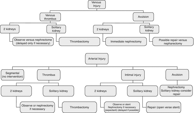
Fig. 21.1
Algorithm for the conservative management of renovascular injuries. Top. Venous injuries. Bottom. Arterial injuries (Santucci and Fisher [23]. With permission of The Journal of Trauma Injury, Infection, and Critical Care)
21.2.6.2 Renal Angiography with Arterial Embolization
There are a few case series that have described the use of angiography with arterial embolization for treatment of renovascular injuries. This option is used when the primary problem is arterial bleeding in a hemodynamically normal patient. In one study, all five patients with renovascular injuries underwent superselective arterial embolization, bleeding was controlled without recurrence, and serum creatinine concentration returned to pre-injury values within 10 days [24]. Similar results were obtained in another series with six patients [25]. The small sample sizes in these and other studies make outcome of this treatment option uncertain, and further studies are needed to define the exact group of renovascular trauma patients that would benefit from this technique.
21.2.6.3 Vascular Repair
Vascular repair is one of the “renal preservation strategies” and is especially indicated if a patient has a solitary kidney or in cases of bilateral renal injuries [7, 26–31]. Repair can be done primarily or by use of a bypass graft. Outcomes following repair are described below subsequently. The majority of authors suggest that success of arterial reconstruction depends on the duration and degree of ischemia and presence or absence of accessory renal arteries providing collateral flow, with irreversible damage occurring when the warm renal ischemia time exceeds 2 h [2, 32]. A lack of relationship between outcome and time to definitive surgery is also reported, because the time of warm ischemia that is tolerated is so short [7]. This is the primary consideration when deciding whether to undertake an interventional or observational approach to renal injury.
21.2.6.4 Partial Nephrectomy/Parenchymal Repair
Partial nephrectomy/parenchymal repair has both been used in the management of renovascular injuries [7]. The segmental vascular supply of the kidney makes this feasible, as one segment can be removed without compromising the functions of the other segments.
21.2.6.5 Immediate (Total) Nephrectomy
Immediate nephrectomy can be used as the primary treatment modality in severe renovascular injury of the grade IV/V variety. It is also indicated when there is failure of vascular repair with either infectious or hypertensive complications resulting from the nonfunctional kidney.
The operative approach to the vasculature of the right kidney usually requires a Cattell-Braasch maneuver (right medial visceral rotation) for exposure of the kidney, renal vessels, and associated right-sided retroperitoneal vascular structures. For the left kidney and vascular structures, a Mattox maneuver (left-sided medial visceral rotation) is usually undertaken. These approaches are used for addressing renal vascular injuries or when a partial nephrectomy is to be done. The primary technical consideration for total nephrectomy is whether vascular control is obtained prior to entering Gerota’s fascia and mobilizing the kidney. From a practical standpoint, this is usually dictated by the patient’s hemodynamic status. In patients with rapidly expanding hematomas, it is usually most expeditious to enter Gerota’s fascia, quickly mobilize the kidney, and control the bleeding with manual pressure or clamps once the kidney and renal vasculature are mobilized. If the patient’s hemodynamic status allows, the approach above to the renal vessels preserves the option of a partial nephrectomy.
21.2.7 Postoperative Management (Including Rehabilitation/Follow-Up)
21.2.7.1 Follow-Up Imaging Studies
These may include abdominal CT scan, angiography, or renal scintigraphy [7]. Renal scintigraphy provides a quantitative assessment of renal function, especially when attempts have been made to salvage the kidneys [7, 33]. The patient with renal dysfunction becomes symptomatic when renal function drops below 25 %. When renal function drops below 10–15 %, dialysis is required to sustain life [34].
21.2.7.2 Serial Measurement of Blood Pressure
Blood pressure monitoring helps to detect the onset of renovascular hypertension. If this occurs, it may be controlled with medication but ultimately often requires nephrectomy.
21.2.7.3 Serum Chemistry
Serial measurement of serum creatinine and electrolytes is important to monitor renal function. However, if the patient has one normal kidney, the serum creatinine may not change even with no function of the contralateral kidney, so this must not be used as the sole measure to follow renal function.
21.2.8 Complications and Pitfalls
21.2.8.1 Renovascular Hypertension
The incidence of posttraumatic renovascular hypertension in the literature ranges from 3.2 to 50 %, with the true incidence reported to be close to about 5 % [7, 37–42]. Several explanations have been proposed to describe the mechanism of hypertension following renovascular trauma, including renal artery stenosis or occlusion resulting from thrombosis, compression, or an intimal flap, as well as the development of a restrictive fibrous capsule around the traumatized kidney that compresses the renal parenchyma and limits normal blood flow [35, 37, 43]. Both of these result in activation of the renin-angiotensin-aldosterone system, with subsequent salt and water retention and widespread vasoconstriction of arterioles. Intractable renovascular hypertension is amenable to nephrectomy, and surgery may be the optimal treatment option [7, 19, 44–47].
21.2.8.2 Diminished Renal Function
Renal dysfunction may be reversible or may progress to acute or chronic renal failure. Serial measurement of serum creatinine is extremely insensitive to changes in renal function of the injured kidney in the presence of a normal contralateral kidney.
21.2.9 Outcomes
Post-injury renal function with poor outcome was defined by Knudson et al. as any of the following [7]:
Renal failure (defined as requiring dialysis or serum creatinine ≥2 mg/dL)
Renal scan showing less than 25 % function of the injured kidney
Post-injury hypertension requiring medication
Delayed nephrectomy, i.e., nephrectomy performed greater than 24 h after injury but during the index hospitalization
Similar clinical outcome measures have also being used by other investigators [34].
Factors affecting outcome include:
Mechanism of Injury: Knudson et al. showed that patients with blunt renovascular injuries were about 2.29 times more likely to have a poor outcome compared to those with penetrating injuries [7].
Type of Vascular Repair: In the same study, for patients with grade IV injuries, outcomes were significantly worse in patients who had arterial repairs compared to those with venous repairs [7]. Other studies have supported this finding [48].
Grade of Renal Injury: For those with grade IV injuries, outcomes were significantly better in those who had a partial nephrectomy or parenchymal repair, compared to those who had a total nephrectomy [7]. For those with grade V injuries, outcomes were significantly better in those who had immediate nephrectomy, compared to those who underwent arterial repair and those who had no renal surgery. In fact, in patients with grade V injuries, attempts at arterial repair or bypass were 15 times more likely to result in a poor outcome, compared with immediate nephrectomy.
Age: Some investigators have reported that immediate nephrectomy (85.7 % good outcomes) and arterial repairs (66.7 % good outcomes) produce the best results in the pediatric population with grades IV and V renal injuries [7]. In this study, 40 % of children treated expectantly, i.e., nonoperatively, had a poor outcome. However, recent evidence suggests that in children with unilateral renovascular injury treated with either nephrectomy or renal preservation, the outcomes were comparable, providing a strong argument for renal preservation [34].
21.3 Ureter
21.3.1 Background/Incidence/Epidemiology
Like renovascular injuries, injuries to the ureters are relatively rare. Injuries to the blood vessels that supply the ureters do not occur in isolation from ureteral injuries themselves. Therefore, a snapshot of ureteral trauma is provided, with specific highlights of vascular considerations as applicable. This chapter focuses on ureteral injury due to external trauma rather than iatrogenic injuries.
The incidence of ureteral injuries from external trauma is estimated to range from less than 1 % to about 2.6 % of all genitourinary injuries, with blunt trauma accounting for about 61.5 % of cases and penetrating trauma accounting for the remaining 38.5 % [49, 50]. Siram et al. reported mortality rates of 6 % for penetrating trauma and 9 % for blunt trauma, although these differences were not statistically significant [49]. In that study, penetrating trauma patients had a higher incidence of vascular injuries (38 %). In patients with penetrating trauma, the most commonly injured vessels were the iliac vein (18 %), iliac artery (10 %), and the inferior vena cava (7 %), whereas, in the blunt trauma subset, the iliac artery (6 %), renal artery (4 %), and renal vein (3 %) were the most commonly injured. Therefore, in the evaluation of any patient with ureteral trauma, a careful search for associated blood vessel injury should be carried out. Alternatively, and more commonly, the surgeon is operating for major vascular injury of the iliac or renal vessels, and after control of the vascular injuries, the surgeon is obliged to evaluate the ureter adjacent to the vascular injury.
21.3.2 Anatomy/Physiology
The ureters are a pair of tubular structures composed largely of smooth muscles that convey urine from the kidneys to the bladder where it is stored prior to micturition. In the adult, the ureter is about 25 cm in length and has a diameter of about 3 mm [51]. The ureter is retroperitoneal, with the upper half located in the abdomen, where it exits the kidney from the renal pedicle, and the lower half located in the pelvis, where it enters the bladder at an oblique angle. The ureter receives multiple blood supplies. The abdominal part is supplied by branches from the renal artery, the abdominal aorta, and the gonadal artery, whereas the pelvic part is supplied by the common iliac artery, internal iliac artery, inferior vesical artery (in the male), or uterine artery (in the female) [51]. The veins draining the ureter generally correspond to the arteries. The peristaltic actions of the ureteral smooth muscles help to actively convey urine from the kidney to the bladder. Reflux in the opposite direction is normally prevented by the oblique angling of the ureter as it enters the bladder.
Stay updated, free articles. Join our Telegram channel

Full access? Get Clinical Tree


