Hypothenar Hammer Syndrome
HARI R. KUMAR and HERON E. RODRIGUEZ
Presentation
A 60-year-old man presents to the emergency department with 10 days of pain and bluish discoloration of the third to fifth digits of his right hand. He is right hand dominant and works as a mechanic. He frequently gets small cuts on his hand and has noticed difficulty healing these wounds on the distal aspects of his fourth and fifth digits. He denies any previous episodes of symptoms or recent major trauma to the extremity. There are no paresthesias in the affected area. The bluish discoloration was not preceded by any other color changes. The patient is a nonsmoker. A hand x-ray is negative for acute fracture.
Differential Diagnosis
Ischemia of the hand has a broad differential diagnosis. The most common etiology is embolization from a proximal source including the heart or upper extremity arterial system. Cardiac sources include emboli from atrial fibrillation or ventricular aneurysm, as well as mycotic emboli from endocarditis. Proximal arterial sources can include plaque from the aortic arch or emboli from a subclavian aneurysm due to thoracic outlet syndrome. Thrombosis from a hypercoagulable state should be considered as well.
Small vessel pathologies can affect the hand and include primary and secondary Raynaud’s phenomenon and thromboangiitis obliterans (Buerger’s disease). Vasculitis causing upper extremity ischemia can be associated with autoimmune or connective tissue disorders, such as scleroderma, systemic lupus erythematosus, rheumatoid arthritis, or Sjögren’s syndrome.
Other causes of hand ischemia include steal syndrome from a proximal arteriovenous fistula/graft, iatrogenic arterial injury, radiation arteritis, injury due to intravenous drug abuse, and occupational repetitive injury. In this latter category, hand-arm vibration syndrome, thenar hammer syndrome, and hypothenar hammer syndrome (HHS) should be considered in manual laborers.
Workup
While the differential for hand ischemia is quite broad, a thorough history and physical can help to narrow the list. The inciting factors for ischemia, such as cold temperature, should be documented. Any changes in the color of the hand, such as the typical pallor, cyanosis, and rubor known as Raynaud’s phenomenon, should be documented. Specific questions should also be asked about the type of work done with hands, including repetitive striking of the palm or use of vibratory tools. The use of tobacco products or intravenous drugs should be documented.
The vascular exam of the hand should include bilateral palpation and auscultation of the brachial, radial, ulnar, and digital arteries and the superficial palmar arch. An Allen’s test should be performed to look for ulnar artery occlusion. Vascular laboratory studies to document digital waveforms and pressures should be obtained.
The chest should be auscultated for abnormal heart sounds or rhythms. Cardiac studies should include an EKG to evaluate for arrhythmia and an echocardiogram to evaluate for sources of embolus.
Laboratory tests for thrombophilia and autoimmune disease should be ordered if hypercoagulability or vasculitis is suspected.
Imaging of the arteries from the aortic arch to the digital arteries should be obtained. Although conventional digital subtraction angiography remains the gold standard, magnetic resonance and computed tomography angiography provides an excellent level of anatomical detail and is the study of choice in many centers. Adequate visualization of the arteries of the hand is essential in operative planning.
Diagnosis
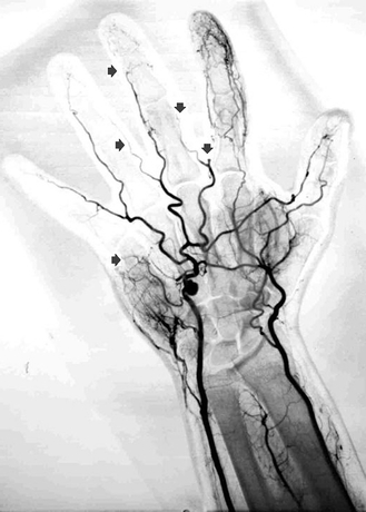
This patient presents with symptoms of hand ischemia and an occupational history of repetitive striking of his hands. He states that in his work as a mechanic, he repeatedly strikes objects with his hand. He is a nonsmoker. He has no history of Raynaud’s or any autoimmune disorder. Physical examination and noninvasive flow showed normal flow to the wrist with evidence of ischemia in the digital arteries of the third to fifth fingers. The Allen’s test is positive. His workup did not demonstrate a proximal embolic source. An angiogram demonstrated embolic occlusion of multiple digital arteries and an ulnar artery aneurysm (Fig. 1). All of these findings are consistent with hypothenar hammer syndrome.

FIGURE 1 Selective arteriogram of the hand showing a saccular aneurysm of the distal portion of the ulnar artery. There is embolic occlusion of multiple digital arteries (arrows).
Discussion
The ulnar artery is vulnerable to injury at the hypothenar eminence. Distal to the wrist, the ulnar artery passes through Guyon’s canal bound by the pisiform and hamate bones. Here, the ulnar artery is in a superficial location and cushioned only by skin, subcutaneous tissue, and the thin palmaris brevis muscle. Repetitive use of the palm of the hand as a “hammer” compresses the unprotected ulnar artery against the nearby hook of the hamate bone, which serves as an “anvil.”
Previous reports of the disease exist, but it was Conn and Bergan who, in 1970, first recognized the anatomic mechanism and coined the term “hypothenar hammer syndrome.” The incidence of the disease is low, with HHS seen in only 1.1% to 1.6% of referrals to tertiary centers for hand ischemia; however, the condition is likely underreported, as Little and Ferguson’s study of 79 mechanics showed 14% had ulnar artery occlusion, but none experienced symptoms severe enough to seek out medical attention.
Both aneurysm formation and occlusive disease can be seen with the disease and depend upon the type of injury to the arterial wall. Damage to the intimal layer can cause vasospasm and platelet aggregation leading to thrombus formation. Damage to the medial layer can cause aneurysm formation, which can also be a nidus for distal emboli. Histologic examination of the resected ulnar artery specimens shows disruption of the internal elastic lamina and hyperplastic proliferation of the intima or media. There is currently debate whether the pathophysiology represents changes from repetitive trauma or if there is a coexistent focal arterial pathology, such as fibromuscular dysplasia to which the repetitive trauma is superimposed.
Patients may present with Raynaud’s phenomenon. However, several features can distinguish HHS from other syndromes. The patient demographic is mostly male manual laborers with a history of repetitive striking of the hand. The dominant hand is the most affected one, and there is a lack of symptoms in the lower extremity. The three fingers on the ulnar side are the most affected. The thumb is usually spared. Cyanosis and pallor can occur, but the hyperemic rubor seen with Raynaud’s phenomenon tends to be absent. A positive Allen’s test, indicating ulnar artery occlusion, can be present. A pulsatile mass overlying the hypothenar eminence can indicate an aneurysm.
Treatment
Patients with mild symptoms and without an aneurysm can be treated nonoperatively. Padded hand protection and avoidance of further repetitive trauma is associated with significant clinical improvement. Tobacco cessation should be recommended if applicable. Medical therapies can include calcium channel blockers, antiplatelet agents, and/or anticoagulation. Thrombolysis can be used in the acute setting in cases of distal embolization. Surgical intervention is indicated in patients with an aneurysm or severe hand ischemia. Surgical intervention can include simple ligation of the aneurysm if there is adequate collateral circulation; typically, resection of the affected segment with reconstruction is required. Previous treatment modalities have included thoracic sympathectomy, in an attempt to increase skin perfusion. The results from this approach have been disappointing.
In the case of this patient who presented with an aneurysm and severe hand ischemia, surgical treatment was recommended. A positive Allen’s test and arterial noninvasive studies consistent with ischemia would preclude simple ligation of the aneurysm. The patient was offered resection with reconstruction.
Surgical Approach
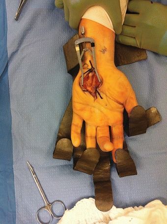
The procedure is performed in the operating room and can be done under general anesthesia or axillary block. The patient is placed in the supine position with arm abducted. The surgical field is prepped and draped in a sterile fashion (Fig. 2).

FIGURE 2 Operative exposure is obtained with the arm abducted and the fingers extended. An incision is made over the hypothenar eminence of the hand just distal to the wrist crease and is carried into the mid palm.


