Hernias

Chapter 46 Hernias


More than 600,000 hernias are repaired annually in the United States, making hernia repair one of the most common operations performed by general surgeons. Despite the frequency of this procedure, no surgeon has ideal results, and complications such as postoperative pain, nerve injury, infection, and recurrence remain.


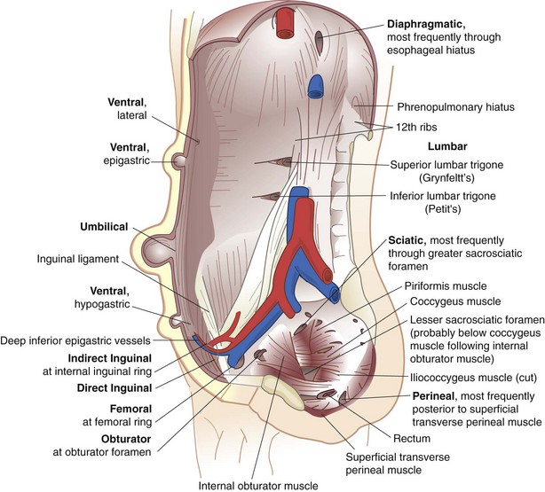
Hernia is derived from the Latin word for rupture. A hernia is defined as an abnormal protrusion of an organ or tissue through a defect in its surrounding walls. Although a hernia can occur at various sites of the body, these defects most commonly involve the abdominal wall, particularly the inguinal region. Abdominal wall hernias occur only at sites at which the aponeurosis and fascia are not covered by striated muscle (Box 46-1). These sites most commonly include the inguinal, femoral, and umbilical areas, linea alba, lower portion of the semilunar line, and sites of prior incisions (Fig. 46-1). The so-called neck or orifice of a hernia is located at the innermost musculoaponeurotic layer, whereas the hernia sac is lined by peritoneum and protrudes from the neck. There is no consistent relationship between the area of a hernia defect and the size of a hernia sac.


A hernia is reducible when its contents can be replaced within the surrounding musculature, and it is irreducible or incarcerated when it cannot be reduced. A strangulated hernia has compromised blood supply to its contents, which is a serious and potentially fatal complication. Strangulation occurs more often in large hernias that have small orifices. In this situation, the small neck of the hernia obstructs arterial blood flow, venous drainage, or both to the contents of the hernia sac. Adhesions between the contents of the hernia and peritoneal lining of the sac can provide a tethering point that entraps the hernia contents and predisposes to intestinal obstruction and strangulation. A more unusual type of strangulation is a Richter’s hernia. In Richter’s hernia, a small portion of the antimesenteric wall of the intestine is trapped within the hernia, and strangulation can occur without the presence of intestinal obstruction.


An external hernia protrudes through all layers of the abdominal wall, whereas an internal hernia is a protrusion of intestine through a defect in the peritoneal cavity. An interparietal hernia occurs when the hernia sac is contained within a musculoaponeurotic layer of the abdominal wall. In broad terms, most abdominal wall hernias can be separated into inguinal and ventral hernias. This chapter focuses on the specific aspects of each of these conditions individually.


Inguinal Hernias


Inguinal hernias are classified as direct or indirect. The sac of an indirect inguinal hernia passes from the internal inguinal ring obliquely toward the external inguinal ring and ultimately into the scrotum. In contrast, the sac of a direct inguinal hernia protrudes outward and forward and is medial to the internal inguinal ring and inferior epigastric vessels. As indirect hernias enlarge, it sometimes can be difficult to distinguish between indirect and direct inguinal hernias. This distinction is of little importance because the operative repair of these types of hernias is similar. A pantaloon-type hernia occurs when there is an indirect and direct hernia component.


Incidence


Hernias are a common problem; however, their true incidence is unknown. It is estimated that 5% of the population will develop an abdominal wall hernia, but the prevalence may be even higher. About 75% of all hernias occur in the inguinal region. Two thirds of these are indirect and the remainder are direct inguinal hernias. Femoral hernias comprise only 3% of all groin hernias.


Men are 25 times more likely to have a groin hernia than women. An indirect inguinal hernia is the most common hernia, regardless of gender. In men, indirect hernias predominate over direct hernias at a ratio of 2 : 1. Direct hernias are uncommon in women. The female-to-male ratio in femoral and umbilical hernias, however, is about 10 : 1 and 2 : 1, respectively. Although femoral hernias occur more frequently in women than in men, inguinal hernias remain the most common hernia in women. Femoral hernias are rare in men. Ten percent of women and 50% of men who have a femoral hernia have or will develop an inguinal hernia.


Indirect inguinal and femoral hernias occur more commonly on the right side. This is attributed to a delay in atrophy of the processus vaginalis after the normal slower descent of the right testis to the scrotum during fetal development. The predominance of right-sided femoral hernias is thought to be caused by the tamponading effect of the sigmoid colon on the left femoral canal.


The prevalence of hernias increases with age, particularly for inguinal, umbilical, and femoral hernias. The likelihood of strangulation and need for hospitalization also increase with aging. Strangulation, the most common serious complication of a hernia, occurs in only 1% to 3% of groin hernias and is more common at the extremes of life. Most strangulated hernias are indirect inguinal hernias; however, femoral hernias have the highest rate of strangulation (15% to 20%) of all hernias and it is therefore recommended that all femoral hernias be repaired at the time of discovery.


Anatomy of the Groin


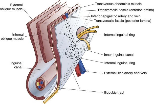
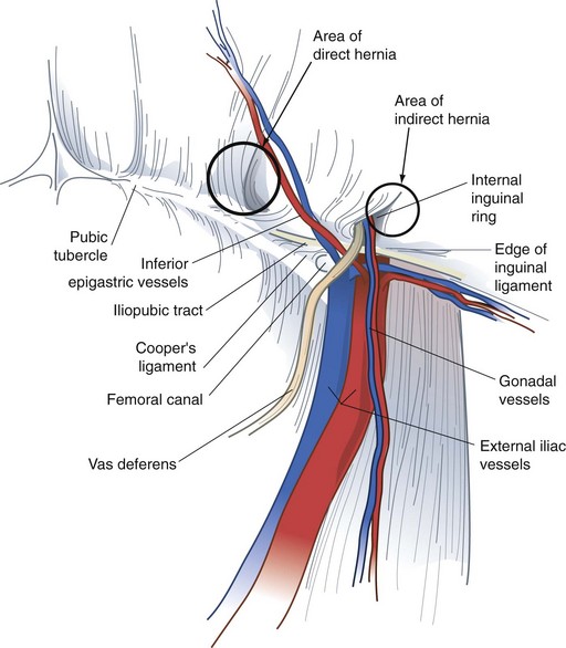
The surgeon must have a comprehensive understanding of the anatomy of the groin to select and use various options for hernia repair properly. In addition, the relationships of muscles, aponeuroses, fascia, nerves, blood vessels, and spermatic cord structures in the inguinal region must be completely understood to obtain the lowest incidence of recurrence and avoid complications. These anatomic considerations must be understood from the anterior and posterior approaches because both are useful in different situations (Figs. 46-2 and 46-3).


From anterior to posterior, the groin anatomy includes the skin and subcutaneous tissues, below which are the superficial circumflex iliac, superficial epigastric, and external pudendal arteries and accompanying veins. These vessels arise from and drain to the proximal femoral artery and vein, respectively, and are directed superiorly. If encountered during operation, these vessels can be retracted or even divided when necessary.


Transversus Abdominis Muscle and Aponeurosis and Transversalis Fascia


The transversus abdominis muscle layer is oriented horizontally throughout most of its area; in the inguinal region, these fibers course in a slightly oblique downward direction. The strength and continuity of this muscle and aponeurosis are important for the prevention and treatment of inguinal hernia.


The aponeurosis of the transversus abdominis covers anterior and posterior surfaces. The lower margin of the transversus abdominis arches along with the internal oblique muscle over the internal inguinal ring to form the transversus abdominis aponeurotic arch. The transversalis fascia is the connective tissue layer that underlies the abdominal wall musculature. The transversalis fascia, sometimes referred to as the endoabdominal fascia, is a component of the inguinal floor. It tends to be denser in this area but still remains relatively thin.


The iliopubic tract is an aponeurotic band that is formed by the transversalis fascia and transversus abdominis aponeurosis and fascia. The iliopubic tract is located posterior to the inguinal ligament and crosses over the femoral vessels and inserts on the anterior superior iliac spine and inner lip of the wing of the ilium.


The inferior crus of the deep inguinal ring is comprised of the iliopubic tract; the superior crus of the deep ring is formed by the transversus abdominis aponeurotic arch. The lateral border of the internal ring is connected to the transversus abdominis muscle, which forms a shutter mechanism to limit the development of an indirect hernia.


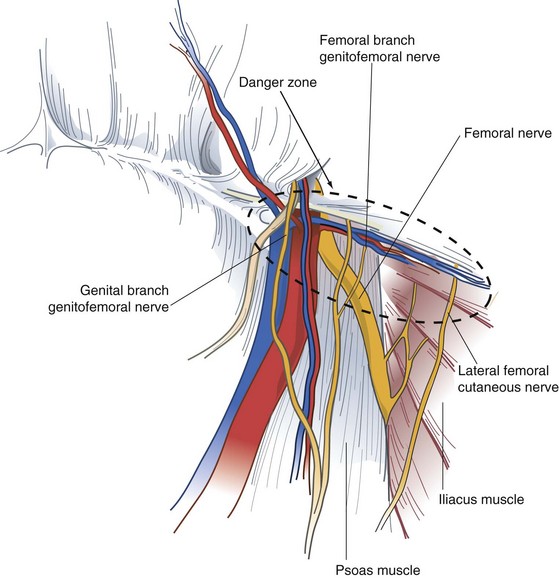
The iliopubic tract is an extremely important structure in the repair of hernias from the anterior and posterior approaches. It comprises the inferior margin of most anterior repairs. The portion of the iliopubic tract lateral to the internal inguinal ring serves as the inferior border below which staples or tacks are not placed during a laparoscopic repair because the femoral, lateral femoral cutaneous, and genitofemoral nerves are located inferior to the iliopubic tract. Although it cannot always be visualized during posterior repairs, if the tacking device cannot be palpated on the anterior abdominal wall, one must assume it is below the iliopubic tract.


Inguinal Canal


The inguinal canal is about 4 cm in length and is located just cephalad to the inguinal ligament. The canal extends between the internal (deep) inguinal and external (superficial) inguinal rings. The inguinal canal contains the spermatic cord in men and the round ligament of the uterus in women.


The spermatic cord is composed of the cremasteric muscle fibers, testicular artery and accompanying veins, genital branch of the genitofemoral nerve, vas deferens, cremasteric vessels, lymphatics, and processus vaginalis. These structures enter the cord at the internal inguinal ring and vessels and vas deferens exit the external inguinal ring. The cremaster muscle arises from the lowermost fibers of the internal oblique muscle and encompasses the spermatic cord in the inguinal canal. The cremasteric vessels are branches of the inferior epigastric vessels and pass through the posterior wall of the inguinal canal through their own foramen. These vessels supply the cremaster muscle and can be divided to expose the floor of the inguinal canal during hernia repair without damaging the testis.


The inguinal canal is bounded superficially by the external oblique aponeurosis. The internal oblique and transversus abdominis musculoaponeuroses form the cephalad wall of the inguinal canal. The inferior wall of the inguinal canal is formed by the inguinal ligament and lacunar ligament. The posterior wall, or floor of the inguinal canal, is formed by the aponeurosis of the transversus abdominis muscle and transversalis fascia.


Hesselbach’s triangle refers to the margins of the floor of the inguinal canal. The inferior epigastric vessels serve as its superolateral border, the rectus sheath as medial border, and the inguinal ligament and pectineal ligament as the inferior border. Direct hernias occur within Hesselbach’s triangle, whereas indirect inguinal hernias arise lateral to the triangle. It is not uncommon, however, for medium and large indirect inguinal hernias to involve the floor of the inguinal canal as they enlarge.


The iliohypogastric and ilioinguinal nerves and genital branch of the genitofemoral nerve are the important sensory nerves in the groin area (Fig. 46-4). The iliohypogastric and ilioinguinal nerves provide sensation to the skin of the groin, base of the penis, and ipsilateral upper medial thigh. The iliohypogastric and ilioinguinal nerves lie beneath the internal oblique muscle to a point just medial and superior to the anterior superior iliac spine, where they penetrate the internal oblique muscle and course beneath the external oblique aponeurosis. The main trunk of the iliohypogastric nerve runs on the anterior surface of the internal oblique muscle and aponeurosis medial and superior to the internal ring. The iliohypogastric nerve may provide an inguinal branch that joins the ilioinguinal nerve. The ilioinguinal nerve runs anterior to the spermatic cord in the inguinal canal and branches at the superficial inguinal ring. The genital branch of the genitofemoral nerve innervates the cremaster muscle and skin on the lateral side of the scrotum and labia. This nerve lies on the iliopubic tract and accompanies the cremaster vessels to form a neurovascular bundle.


Preperitoneal Space


The preperitoneal space contains adipose tissue, lymphatics, blood vessels, and nerves. The nerves of the preperitoneal space of specific concern to the surgeon include the lateral femoral cutaneous nerve and genitofemoral nerve. The lateral femoral cutaneous nerve originates as a root of L2 and L3 and is occasionally a direct branch of the femoral nerve. This nerve courses along the anterior surface of the iliac muscle beneath the iliac fascia and passes under or through the lateral attachment of the inguinal ligament at the anterior superior iliac spine. This nerve runs beneath or occasionally through the iliopubic tract, lateral to the internal inguinal ring.


The genitofemoral nerve usually arises from the L2 or L1-L2 nerve roots. It divides into genital and femoral branches on the anterior surface of the psoas muscle. The genital branch enters the inguinal canal through the deep ring, whereas the femoral branch enters the femoral sheath lateral to the artery.


The inferior epigastric artery and vein are branches of the external iliac vessels and are important landmarks for laparoscopic hernia repair. These vessels course medial to the internal inguinal ring and eventually lie beneath the rectus abdominis muscle, immediately superficial to the transversalis fascia. The inferior epigastric vessels serve to define the types of inguinal hernia. Indirect inguinal hernias occur lateral to the inferior epigastric vessels, whereas direct hernias occur medial to these vessels.


The deep circumflex iliac artery and vein are located below the lateral portion of the iliopubic tract in the preperitoneal space. These vessels are branches of the inferior epigastric or external iliac artery and vein. It is important to dissect only above the iliopubic tract during a laparoscopic hernia repair to avoid injury to these vessels.


The vas deferens courses through the preperitoneal space from caudad to cephalad and medial to lateral to join the spermatic cord at the deep inguinal ring.


Diagnosis


A bulge in the inguinal region is the main diagnostic finding in most groin hernias. There may be associated pain or vague discomfort in the region, but groin hernias are usually not extremely painful unless incarceration or strangulation has occurred. In the absence of physical findings, alternative causes for pain need to be considered. Occasionally, patients may experience paresthesias related to compression or irritation of the inguinal nerves by the hernia. Masses other than hernias can occur in the groin region. Physical examination alone often differentiates between a groin hernia and these masses (Box 46-2).


The inguinal region is examined with the patient in the supine and standing positions. The examiner visually inspects and palpates the inguinal region, looking for asymmetry, bulges, or a mass. Having the patient cough or perform a Valsalva maneuver can facilitate identification of a hernia. The examiner places a fingertip over the inguinal canal and repeats the examination. Finally, a fingertip is placed into the external inguinal ring by invaginating the scrotum to detect a small hernia. A bulge moving lateral to medial in the inguinal canal suggests an indirect hernia. If a bulge progresses from deep to superficial through the inguinal floor, a direct hernia is suspected. This distinction is not critical because repair is approached the same way, regardless of the type of hernia. A bulge identified below the inguinal ligament is consistent with a femoral hernia.


A bulge of the groin described by the patient that is not demonstrated on examination presents a dilemma. Having the patient stand or ambulate for a period of time may allow an undiagnosed hernia to become visible or palpable. If a hernia is strongly suspected, but undetectable, a repeat examination at another time may be helpful.


Ultrasonography also can aid in the diagnosis. There is a high degree of sensitivity and specificity for ultrasound in the detection of occult direct, indirect, and femoral hernias.1 Other imaging modalities are less useful. Computed tomography (CT) of the abdomen and pelvis may be useful for the diagnosis of obscure and unusual hernias as well as atypical groin masses.2 Occasionally, laparoscopy can be diagnostic and therapeutic for particularly challenging cases.


Treatment


Nonoperative Management


Most surgeons recommend operation on discovery of a symptomatic inguinal hernia because the natural history of a groin hernia is that of progressive enlargement and weakening, with a small potential for incarceration and strangulation. However, in patients with minimal symptoms, the clinician is often faced with balancing the risk for hernia-related complications such as incarceration and bowel strangulation, with the potential for complications in the short and long term. Fitzgibbons and colleagues3 have reported a prospective randomized trial of a watchful waiting strategy for men with asymptomatic or minimally symptomatic inguinal hernias. These investigators randomized more than 700 men to a watchful waiting or open tension-free hernia repair. At 2 years of follow-up, there were no deaths attributed to the study and the risk for hernia incarceration in the watchful waiting group was extremely low, 0.3% of study participants or 1.8 events/1000 patient-years. Almost 25% of patients assigned to watchful waiting crossed over to the surgical group, usually for pain related to the hernia that limited activity. Despite the seemingly high crossover rate, those patients, who later had surgery, did not have increased surgical site infections, longer operative times, or higher recurrence rates than those who were initially assigned to early repair. This study provides conclusive evidence that a strategy of watchful waiting is safe for older patients with asymptomatic or minimally symptomatic inguinal hernias and that even though almost 25% of patients eventually undergo repair, when they do, the operative risks and complication rates are no different than those of patients undergoing prophylactic repair. Watchful waiting also is a cost-effective management strategy for patients with no or minimal symptoms.


Patients electing nonoperative management can occasionally have symptomatic improvement with the use of a truss. This approach is more commonly used in Europe. Spring trusses are more versatile than elastic ones, although most information on their use has been anecdotal. Correct measurement and fitting are important. Hernia control has been reported in about 30% of patients. Complications associated with the use of a truss include testicular atrophy, ilioinguinal or femoral neuritis, and hernia incarceration.


It is generally agreed that nonoperative management is not used for femoral hernias because of the high incidence of associated complications, particularly strangulation.


Operative Repair


Anterior Repairs


Anterior repairs are the most common operative approach for inguinal hernias. Tension-free repairs are now standard and there are a variety of different types. Older tissue types of repair are rarely indicated, except for patients with simultaneous contamination or concomitant bowel resection, when placement of a mesh prosthesis may be contraindicated.


There are some technical aspects of the operation common to all anterior repairs. Open hernia repair is begun by making a transversely oriented linear or slightly curvilinear incision above the inguinal ligament and a fingerbreadth below the internal inguinal ring. The internal inguinal ring is located topographically at the midpoint between the anterior superior iliac spine and ipsilateral pubic tubercle. Dissection is continued through the subcutaneous tissues and Scarpa’s fascia. The external oblique fascia and external inguinal ring are identified. The external oblique fascia is incised through the superficial inguinal ring to expose the inguinal canal. The genital branch of the genitofemoral nerve, as well as the ilioinguinal and iliohypogastric nerves, are identified and avoided or mobilized to prevent transection and entrapment. The spermatic cord is mobilized at the pubic tubercle by a combination of blunt and sharp dissection. Improper mobilization of the spermatic cord too lateral to the pubic tubercle can cause confusion in the identification of tissue planes and essential structures, and may result in injury to the spermatic core structures or disruption of the floor of the inguinal canal.


The cremasteric muscle of the mobilized spermatic cord is separated parallel to its fibers from the underlying cord structures. The cremasteric artery and vein, which join the cremaster muscle near the inguinal ring, can usually be avoided but may need to be cauterized or ligated and divided. When an indirect hernia is present, the hernia sac is located deep to the cremaster muscle and anterior and superior to the spermatic cord structures. Incising the cremaster muscle in a longitudinal direction and dividing it circumferentially near the internal inguinal ring help expose the indirect hernia sac. The hernia sac is carefully separated from adjacent cord structures and dissected to the level of the internal inguinal ring. The sac is opened and examined for visceral contents if it is large; however, this step is unnecessary in small hernias. The sac can be mobilized and placed within the preperitoneal space, or the neck of the sac can be ligated at the level of the internal ring and any excess sac excised. If a large hernia sac is present, it can be divided using electrocautery to facilitate ligation. It is not necessary to excise the distal portion of the sac. If the sac is broad-based, it may be easier to displace it into the peritoneal cavity rather than ligate it. Direct hernia sacs protrude through the floor of the inguinal canal and can be reduced below the transversalis fascia before repair. A lipoma of the cord actually represents retroperitoneal fat that has herniated through the deep inguinal ring; this should be suture-ligated and removed.


A sliding hernia presents a special challenge in handling the hernia sac. With a sliding hernia, a portion of the sac is composed of visceral peritoneum covering part of a retroperitoneal organ, usually the colon or bladder. In this situation, the grossly redundant portion of the sac (if present) is excised and the peritoneum reclosed. The organ and sac then can be reduced below the transversalis fascia, similar to the procedure for a direct hernia.


Tissue Repairs


Although tissue repairs have largely been abandoned because of unacceptably high recurrence rates, they remain useful in certain situations. In strangulated hernias, for which bowel resection is necessary, mesh prostheses are contraindicated and a tissue repair is necessary. Available options for tissue repair include iliopubic tract, Shouldice, Bassini, and McVay repairs.


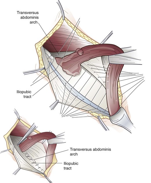
The iliopubic tract repair approximates the transversus abdominis aponeurotic arch to the iliopubic tract with the use of interrupted sutures (Fig. 46-5). The repair begins at the pubic tubercle and extends laterally past the internal inguinal ring. This repair was initially described using a relaxing incision (see later); however, many surgeons who use this repair do not perform a relaxing incision.


The Shouldice repair emphasizes a multilayer imbricated repair of the posterior wall of the inguinal canal with a continuous running suture technique. After completion of the dissection, the posterior wall of the inguinal canal is reconstructed by superimposing running suture lines progressing from deep to more superficial layers. The initial suture line secures the transversus abdominis aponeurotic arch to the iliopubic tract. Next, the internal oblique and transversus abdominis muscles and aponeuroses are sutured to the inguinal ligament. The Shouldice repair is associated with a very low recurrence rate and a high degree of patient satisfaction in highly selected patients.


The Bassini repair is performed by suturing the transversus abdominis and internal oblique musculoaponeurotic arches or conjoined tendon (when present) to the inguinal ligament. This once popular technique is the basic approach to nonanatomic hernia repairs and was the most popular type of repair done before the advent of tension-free repairs.


Cooper’s ligament repair, also known as the McVay repair, has traditionally been popular for the correction of direct inguinal hernias, large indirect hernias, recurrent hernias, and femoral hernias. Interrupted nonabsorbable sutures are used to approximate the edge of the transversus abdominis aponeurosis to Cooper’s ligament. When the medial aspect of the femoral canal is reached, a transition suture is placed to incorporate Cooper’s ligament and the iliopubic tract. Lateral to this transition stitch, the transversus abdominis aponeurosis is secured to the iliopubic tract. An important principle of this repair is the need for a relaxing incision. This incision is made by reflecting the external oblique aponeurosis cephalad and medial to expose the anterior rectus sheath. An incision is then made in a curvilinear direction, beginning 1 cm above the pubic tubercle throughout the extent of the anterior sheath to near its lateral border. This relieves tension on the suture line and results in decreased postoperative pain and hernia recurrence. The fascial defect is covered by the body of the rectus muscle, which prevents herniation at the relaxing incision site. The McVay repair is particularly suited for strangulated femoral hernias because it provides obliteration of the femoral space without the use of mesh.


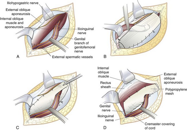
Tension-Free Anterior Inguinal Hernia Repair


The tension-free repair has become the dominant method of inguinal hernia repair (Fig. 46-6). Recognizing that tension in a repair is the principal cause of recurrence, current practices in hernia management use a synthetic mesh prosthesis to bridge the defect, a concept popularized by Lichtenstein. There are several options for placement of mesh during anterior inguinal herniorrhaphy, including the Lichtenstein approach, plug and patch technique, and sandwich technique, with both an anterior and preperitoneal piece of mesh.


image

FIGURE 46-6 Lichtenstein tension-free hernia repair. A, This procedure is performed by careful dissection of the inguinal canal. High ligation of an indirect hernia sac is performed and the spermatic cord structures are retracted inferiorly. The external oblique aponeurosis is separated from the underlying internal oblique muscle high enough to accommodate a 6- to 8-cm-wide mesh patch. Overlap of the internal oblique muscle edge by 2 to 3 cm is necessary. A sheet of polypropylene mesh is fashioned to fit the inguinal canal. A slit is made in the lateral aspect of the mesh and the spermatic cord is placed between the two tails of the mesh. B, The spermatic cord is retracted in the cephalad direction. The medial aspect of the mesh overlaps the pubic bone by approximately 2 cm. The mesh is secured to the aponeurotic tissue overlying the pubic tubercle using a running suture of nonabsorbable monofilament material. The suture is continued laterally by suturing the inferior edge of the mesh to the shelving edge of the inguinal ligament to a point just lateral to the internal inguinal ring. C, A second monofilament suture is placed at the level of the pubic tubercle and continued laterally by suturing the mesh to the internal oblique aponeurosis or muscle approximately 2 cm from the aponeurotic edge. D, The lower edges of the two tails are sutured to the shelving edge of the inguinal ligament to create a new internal ring made of mesh. The spermatic cord structures are placed within the inguinal canal overlying the mesh. The external oblique aponeurosis is closed over the spermatic cord.


(From Arregui ME, Nagan RD [eds]: Inguinal hernia: Advances or controversies? Oxford, England, 1994, Radcliffe Medical.)


In the Lichtenstein repair,4 a piece of prosthetic nonabsorbable mesh is fashioned to fit the canal. A slit is cut into the distal lateral edge of the mesh to accommodate the spermatic cord. There are various preformed, commercially available prostheses available for use. Monofilament nonabsorbable suture is used to secure the mesh, beginning at the pubic tubercle and running a length of suture in both directions toward the superior aspect above the internal inguinal ring to the level of the tails of the mesh. The mesh is sutured to the aponeurotic tissue overlying the pubic bone medially, continuing superiorly along the transversus abdominis or conjoined tendon. The inferolateral edge of the mesh is sutured to the iliopubic tract or shelving edge of the inguinal ligament to a point lateral to the internal inguinal ring. At this point, the tails created by the slit are sutured together around the spermatic cord, snugly forming a new internal inguinal ring. It is important to protect the ilioinguinal nerve and genital branch of the genitofemoral nerve from entrapment by placing them with the cord structures as they are passed through this newly fashioned internal inguinal ring or avoiding their enclosure in the repair.


Adapting the principles of tension-free repair, Gilbert5 has reported using a cone-shaped plug of polypropylene mesh that when inserted into the internal inguinal ring, would deploy like an upside-down umbrella and occlude the hernia. This plug is sewn to the surrounding tissues and held in place by an additional overlying mesh patch. This patch may not need to be secured by sutures; however, to do so requires dissection to create a sufficient space between the external and internal oblique muscles for the patch to lie flat over the inguinal canal. This so-called plug and patch repair, an extension of Lichtenstein’s original mesh repair, has now become the most commonly performed primary anterior inguinal hernia repair. Although this repair can be done without suture fixation by some experienced surgeons, most secure plug and patch with several monofilament nonabsorbable sutures, especially for very weak inguinal floors or large defects.


The sandwich technique involves a bilayered device, with three polypropylene components. An underlay patch provides a posterior repair similar to that of the laparoscopic approach, a connector functions similar to a plug, and an onlay patch covers the posterior inguinal floor. The use of interrupted fixating sutures is not mandatory, but most surgeons place three or four fixation sutures in this repair.


Another option for a tension-free mesh repair involves a preperitoneal approach using a self-expanding polypropylene patch.6 A pocket is created in the preperitoneal space by blunt dissection and then a preformed mesh patch is inserted into the hernia defect, which expands to cover the direct, indirect, and femoral spaces. The patch lies parallel to the inguinal ligament. It can remain without suture fixation, or a tacking suture can be placed.


The Stoppa-Rives repair uses a subumbilical midline incision to place a large mesh prosthesis into the preperitoneal space.7 Blunt dissection is used to create an extraperitoneal space that extends into the prevesical space, beyond the obturator foramen, and posterolateral to the pelvic brim. This technique has the advantage of distributing the natural intra-abdominal pressure across a broad area to retain the mesh in a proper location. The Stoppa-Rives technique is particularly useful for large, recurrent, or bilateral hernias.


Preperitoneal Repair


The open preperitoneal approach is useful for the repair of recurrent inguinal hernias, sliding hernias, femoral hernias, and some strangulated hernias.8 A transverse skin incision is made 2 cm above the internal inguinal ring and is directed to the medial border of the rectus sheath. The muscles of the anterior abdominal wall are incised transversely and the preperitoneal space is identified. If further exposure is needed, the anterior rectus sheath can be incised and the rectus muscle retracted medially. The preperitoneal tissues are retracted cephalad to visualize the posterior inguinal wall and the site of herniation. The inferior epigastric artery and veins are generally beneath the midportion of the posterior rectus sheath and usually do not need to be divided. This approach avoids mobilization of the spermatic cord and injury to the sensory nerves of the inguinal canal, which is particularly important for hernias previously repaired through an anterior approach. If the peritoneum is incised, it is sutured closed to avoid the evisceration of intraperitoneal contents into the operative field. The transversalis fascia and transversus abdominis aponeurosis are identified and sutured to the iliopubic tract with permanent sutures. Femoral hernias repaired by this approach require closure of the femoral canal by securing the repair to Cooper’s ligament. A mesh prosthesis is frequently used to obliterate the defect in the femoral canal, particularly with large hernias.


Laparoscopic Repair


Laparoscopic inguinal hernia repair is another method of tension-free mesh repair, based on a preperitoneal approach. The laparoscopic approach provides the mechanical advantage of placing a large piece of mesh behind the defect covering the myopectineal orifice and using the natural forces of the abdominal wall to support the mesh in place. Proponents have touted quicker recovery, less pain, better visualization of anatomy, usefulness for fixing all inguinal hernia defects, and decreased surgical site infections. Critics have emphasized longer operative times, technical challenges, risk of recurrence, and increased cost. Although controversy exists about the usefulness of laparoscopic repair for primary unilateral inguinal hernias, most agree that this approach has advantages for patients with bilateral or recurrent hernias.9 Adopting practice guidelines for the performance of laparoscopic hernia repairs could help control costs.


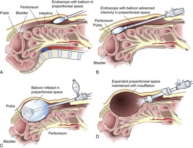
When considering the laparoscopic approach for repair of inguinal hernias, the surgeon has several options. Initially, laparoscopic repairs involved placing a large piece of mesh in an intraperitoneal position, similar to a laparoscopic ventral hernia repair. This approach has been abandoned because of high recurrence rates and the drawbacks of intraperitoneal mesh. The most popular techniques include a totally extraperitoneal (TEP) and transabdominal preperitoneal (TAPP) approach. The main difference between these two techniques is the sequence of gaining access to the preperitoneal space. In the TEP approach, the dissection begins in the preperitoneal space using a balloon dissector. With the TAPP repair, the preperitoneal space is accessed after initially entering the peritoneal cavity. Each approach has its merits. Using the TEP approach, the preperitoneal dissection is quicker and the potential risk for intraperitoneal visceral damage is minimized. However, the use of dissection balloons is costly, the working space is more limited, and it may not be possible to create a working space if the patient has had a prior preperitoneal operation. Also, if a large tear in the peritoneum is created during a TEP approach, the potential working space can become obliterated, necessitating conversion to a TAPP approach. For these reasons, knowledge of the transabdominal technique is essential when performing laparoscopic inguinal hernia repairs. The transabdominal approach allows identification of the groin anatomy before extensive dissection and disruption of natural tissue planes. The larger working space of the peritoneal cavity can make early experience with the laparoscopic approach easier.


There are no absolute contraindications to laparoscopic inguinal hernia repair other than the patient’s inability to tolerate general anesthesia. Patients who have had extensive prior lower abdominal surgery can require significant adhesiolysis and may be best approached anteriorly. In particular, patients who have had a radical retropubic prostatectomy with the preperitoneal space previously dissected can make accurate and safe dissection challenging.


In the TEP approach, an infraumbilical incision is used. The anterior rectus sheath is incised, the ipsilateral rectus abdominis muscle is retracted laterally, and blunt dissection is used to create a space beneath the rectus. A dissecting balloon is inserted deep to the posterior rectus sheath, advanced to the pubic symphysis, and inflated under direct laparoscopic vision (Fig. 46-7). After it is opened, the space is insufflated and additional trocars are placed. A 30-degree laparoscope provides the best visualization of the inguinal region (see Fig. 46-3). The inferior epigastric vessels are identified along the lower portion of the rectus muscle and serve as a useful landmark. Cooper’s ligament must be cleared from the pubic symphysis medially to the level of the external iliac vein. The iliopubic tract is also identified. Care must be taken to avoid injury to the femoral branch of the genitofemoral nerve and lateral femoral cutaneous nerve, which are located lateral to and below the iliopubic tract (see Fig. 46-4). Lateral dissection is carried out to the anterior superior iliac spine. Finally, the spermatic cord is skeletonized.


< div class='tao-gold-member'>

Stay updated, free articles. Join our Telegram channel

Aug 1, 2016 | Posted by in CARDIAC SURGERY | Comments Off on Hernias

Full access? Get Clinical Tree

Get Clinical Tree app for offline access