(1)
Department of General surgery, Fuwai Hospital, Beijing, China
10.1 General Considerations
The primitive cardiovascular system begins forming during the fourth week of human embryonic development. The heart begins to beat, and blood begins to circulate through the primitive heart tube. The blood originates from the cephalic dorsal aorta and flows back to the heart tube through the common cardinal vein. The tube-like primitive heart tube will develop into the complete cardiovascular system step by step. Abnormity during the development of the embryo circulatory system leads to various cardiovascular abnormalities.
The primitive heart tube gradually develops into three layers: endocardium, myocardium, and epicardium. The pericardial sac originates from the cranial end of the primitive heart tube. After the cranial end folds toward the ventral side, the pericardial sac approaches that side of the heart tube, after which the pericardium surrounds the heart tube from the ventral to the dorsal side. Hence, the pericardial reflection is located on the dorsal side around the proximal portion of the main vessels. A more detailed description of the development of the primitive heart is provided in the discussion below.
The development from the primitive heart tube to the mature heart includes four aspects. The first aspect is the determination of heart chambers and the roots of the main arteries. This process includes the elongation of the primitive heart tube, segmentation, and looping. After all of these developmental processes are completed, the three-dimensional structure of the heart is formed. Abnormal determination of the heart does not always result in abnormal blood flow. For example, when malposition of the great arteries occurs, the AV lies in front of the pulmonary artery but the aorta still can receive blood flow from the LV as normal. The second aspect involves the connections of the heart chambers and the roots of the main arteries. If abnormalities in these connections result in an abnormal circulating system, an active correction is required. The third aspect is the septation of the heart chambers and the roots of the main arteries. This process includes the septations of the conus arteriosus, the ventricles, the atrioventricular canal, and the atria. The fourth aspect involves the maturation of the heart chambers, which includes the enlargement of the heart chambers, the formation of the heart valves, and the development of the cardiac conducting system.
10.2 Heart Chambers and Roots of Main Arteries
10.2.1 Formation of the Primitive Heart Tube
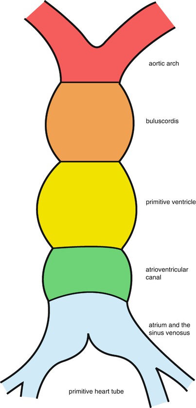
The primitive cardiac cells (cardiac progenitors) develop to form a primitive single chamber, the linear heart tube. The cranial end of the primitive heart tube is called the truncus arteriosus (TA). The primitive ventricle is caudal to the bulbus cordis and the vitelline veins (from the yolk sac). As described above, the pericardial sac moves from the caudal side to the dorsal side of the primitive heart tube and further develops to enclose the primitive heart tube. The pericardial reflex line lies outside the primitive heart tube (Fig. 10.1).


Fig. 10.1
Primitive heart tube. The primitive heart tube initially is bulged and segmented. The bulbus cordis develops into the truncus and conus, and the AV canal is shortened into the atrioventricular foramen. The sinus venosus is merged into the atrium
10.2.2 Segmentation of the Primitive Heart Tube
The primitive heart tube elongates and loops in the pericardial sac and develops many constrictions outlining future structures. The cranial most area is the bulbus cordis, and the ventricles are caudal to it. The AV canal is the caudal-most structure of the tubular heart. The atrium and sinus venosus are not formed yet; they only connect to the AV canal and the vitelline veins. The primitive atrium and sinus venosus lie outside the caudal end of the pericardial sac.
In the development of the primitive heart tube, the bulbus cordis divides into two segments: the truncus, or front, side and the conus, or posterior, side. These two segments together form the conotruncus. The primitive atrium and sinus venosus merge into one chamber, which forms a part of the primitive heart tube. The primitive heart tube is divided into six segments at this point: conus, conotruncus, ventricles, AV canal, atrium, and sinus venosus. The atrium and sinus venosus move upside into the pericardial sac. The conotruncus connects to the first aorta arch at the cepharal end, and the sinus venosus receives the umbilical veins, the vitelline veins, and the common cardinal veins at the caudal end.
10.2.3 Looping of the Primitive Heart Tube
The heart tube elongates and bends in the pericardial sac. This looping process occurs in three directions: right, caudal (posterior), and ventral.
10.2.3.1 Right Direction
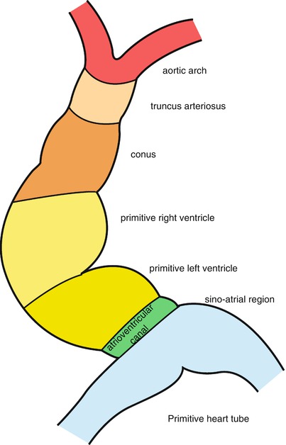
The ventricular segment of the bulbus cordis bends to the right to form the bulboventricular loop. The most extensive bending of the ventricle and the bulbus cordis occurs here as the tube forms a convex curve to the right front side. The internal chamber of this connection is called the ostium bulbi, which functions as the outlet of the primitive ventricle. The cepharal end of the ostium bulbi is the conus, which develops into the outlet of the ventricles. The caudal end of the ostium bulbi develops into the RV. The D-looping process of the bulboventricular loop places the ventricles of the mature heart in the right positions.
10.2.3.2 Caudal Direction
As the bulbus cordis bends to the right, the atrium and the sinus venosus at the caudal end of the AV canal bend to the dorsal side of the ventricles.
10.2.3.3 Ventral Direction
The atrium and the sinus venosus move to the cepharal end and then to the dorsal end of the conotruncus (backward of the ventricles), where the atrium of the mature heart localizes. During this upside bending process, the atrium and the sinus venosus elongate, enlarge, and, for the most part, merge into the pericardium.
After the bending and movement are completed, the heart has a smooth lining except for the area just proximal and just distal to the bulboventricular foramen, where trabeculae form. The primitive ventricle will eventually develop into the LV, and the proximal portion of the bulbus cordis will form the RV. The distal part of the bulbus cordis, an elongated structure, will form the outflow tracts of both ventricles, and the truncus arteriosus will form the roots of both great vessels. The bulbus cordis gradually acquires a more medial position due to the growth of the RA, forcing the bulbus to be in the sulcus between the two atria (Fig. 10.2).


Fig. 10.2
Bending and D-looping of the heart tube. The conus and ventricles elongate and bend to the right, which is called D–looping. The ostium bulbi moves to the right side, and the primitive ventricle lies below. The middle part of the primitive ventricle sprouts into the interventricular ostium, which divides the left and right ventricles. The primitive atrium moves backward and to the upside
10.2.4 Rotation of the Proximal End of the Conotruncus
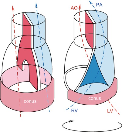
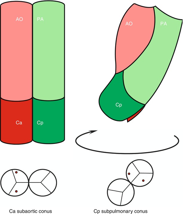
The conotruncus septation divides the conotruncus into two parallel chambers. The distal end of the conotruncus connects to the great arteries; the main artery on the right side becomes the aorta, and the main artery on the left side develops into the pulmonary artery. The proximal end of the conotruncus is the ostium bulbi, and this structure rotates clockwise approximately 90°–110° along the longitudinal axis (Fig. 10.3). At this point, the distal end of the conotruncus and the aortic arch are relatively stable. Due to this rotation, the previous parallel blood flow becomes spiral blood flow. The conotruncus below the aortic valve rotates to the left rear, and the conotruncus below the pulmonary valve rotates to the right front. Concomitantly, the conotruncus itself becomes shortened and is absorbed, with most of the portion below the aortic valve disappearing (Fig. 10.4).


Fig. 10.3
Rotation of the proximal end of the conotruncus. The distal end of the conotruncus septation is relatively stationary, whereas the proximal end rotates. The aortic and the pulmonary valves also rotate: the AV to the left lower rear and the PV to the right upside. Along with the rotation of the conotruncus, the conotruncus below the AV is absorbed. The conotruncus below the PV is also shortened but maintains its structure. AO ascending aorta, PA pulmonary artery, RV right ventricle, LV left ventricle

Fig. 10.4
Rotation and absorption of the proximal end of the conotruncus. At first, the conotruncus is divided into two parallel chambers. The aorta and the subaortic conus (Ca) are located on the right side, whereas the pulmonary artery and the subpulmonary conus (Cp) are on the left side. Then, the aorta and the pulmonary artery remain stationary as the conus rotates. The conotruncus below the AV is absorbed at the same time. AO ascending aorta, PA pulmonary artery
< div class='tao-gold-member'>
Only gold members can continue reading. Log In or Register to continue
Stay updated, free articles. Join our Telegram channel

Full access? Get Clinical Tree


