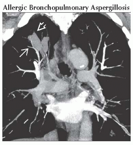
Finger in Glove Appearance
Robert B. Carr, MD
DIFFERENTIAL DIAGNOSIS
Common
Allergic Bronchopulmonary Aspergillosis
Congenital Bronchial Atresia
Less Common
Cystic Fibrosis
Obstructing Mass
ESSENTIAL INFORMATION
Key Differential Diagnosis Issues
Finger in glove refers to mucoid impaction with dilation of large bronchi resulting in tubular or branching opacities
Initially described in patients with allergic bronchopulmonary aspergillosis (ABPA)
Tubular opacities seen on radiography may be bronchial or vascular in origin
Can be confused with arteriovenous malformation on radiography
CT reveals low-attenuation mucus in dilated central bronchi, thus differentiating from vascular causes
Helpful Clues for Common Diagnoses
Allergic Bronchopulmonary Aspergillosis
Hypersensitivity reaction to Aspergillus antigens, usually A. fumigatus
Usually occurs in patients with asthma or cystic fibrosis
Associated with elevated IgE levels and peripheral eosinophilia
Usually affects upper lobes
Secretions may be hyperattenuating on CT due to presence of calcium oxalate
Congenital Bronchial Atresia
Due to congenital atresia of segmental bronchus
Usually incidental finding but may cause recurrent infections in 20% of patients
Most common in apicoposterior segment of left upper lobe, followed by right upper lobe
CT reveals surrounding pulmonary hyperlucency due to air-trapping and oligemia
Helpful Clues for Less Common Diagnoses
Cystic Fibrosis
Congenital disease caused by chloride channel mutation on chromosome 7
Recurrent infections lead to progressive bronchial wall injury
CT findings include bronchiectasis, mucoid impaction, peribronchial thickening, mosaic perfusion, and tree in bud opacities
Obstructing Mass
Rare benign endobronchial tumors include lipoma, papilloma, and hamartoma
Malignant tumors are more common, including carcinoid, bronchogenic carcinoma, and endobronchial metastasis
Obstructing tumor may be directly visualized on CT
Image Gallery
 Coronal CECT MIP image shows finger in glove formation in the right upper lobe bronchi
 . Notice how much larger the bronchi are than the adjacent pulmonary vessels . Notice how much larger the bronchi are than the adjacent pulmonary vessels  . .Stay updated, free articles. Join our Telegram channel
Full access? Get Clinical Tree
 Get Clinical Tree app for offline access

|