Extracranial Carotid Artery Aneurysms


Extracranial Carotid Artery Aneurysms


James C. Stanley


Extracranial carotid artery aneurysms represent an important but uncommon disease. Certain of these aneurysms are associated with thromboembolic complications causing stroke. Rupture of an aneurysm, especially in the case of a false aneurysm following carotid artery endarterectomy, represents a surgical emergency. Clinical manifestations and treatment vary, depending upon the type, size, and location of the aneurysm, warranting individual discussion of true aneurysms, traumatic false aneurysms, and false aneurysms following carotid artery endarterectomy.


True Carotid Artery Aneurysms


Incidence


True aneurysms account for a little more than half of all reported extracranial carotid artery aneurysms. These aneurysms affect men twice as often as women. The average age at recognition is 60 years. Although most true carotid aneurysms exhibit arteriosclerosis, arteriosclerosis is considered a secondary event rather than a primary etiologic factor.


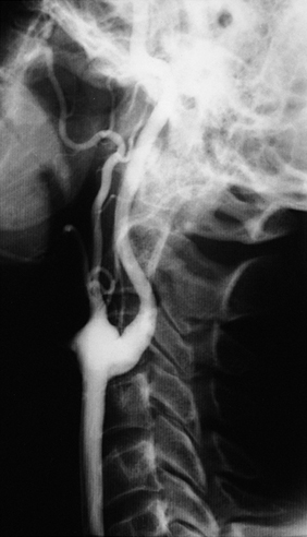
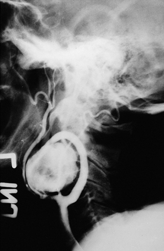
The carotid artery bulb is the site of true aneurysms in more than 95% of cases (Figures 1 and 2). Bilateral aneurysms are noted in 12% of cases, and no predilection exists for right- or left-sided involvement in patients with unilateral aneurysms. Because the normal carotid bulb is usually 40% greater in diameter than the more distal internal carotid artery, it requires very little expansion to reach the 50% dilation level. The latter is a generally accepted definition of an aneurysm. However, given the normal dilation of the carotid bulb, it becomes difficult to define the natural history of borderline-sized aneurysms. Because of concerns over one’s ability to differentiate a generous-sized carotid bulb from a small aneurysm, it has been proposed that an aneurysm only exists when the carotid bulb dilation at least is 200% greater than the extracranial internal carotid artery diameter or 150% of the common carotid artery diameter. Although true arteriosclerotic aneurysms have been encountered in the common carotid artery and the more distal internal carotid artery, trauma or some other vascular insult has likely preceded aneurysm development at these latter sites. Aneurysms affecting other arteries in patients with carotid artery aneurysms have been observed in nearly 25% of cases.


Manifestations


The natural history of true carotid artery aneurysms is poorly defined. Most aneurysms are likely to be asymptomatic, although the surgical literature does not support such a statement. Among patients described in surgical reports, approximately 60% have experienced transient ischemic attacks, including 33% with amaurosis and 21% with hemispheric symptoms. Frank strokes affected an additional 8% of patients.


Rupture of a true carotid aneurysm is rare, having been reported in fewer than a dozen patients. However, acute expansion of true carotid artery aneurysms does occur, and it can cause secondary cranial nerve dysfunction, such as hoarseness related to pressure on the recurrent laryngeal nerve. Severe hemicrania, often with Horner’s syndrome, can accompany acute expansion of an aneurysm, although such is more often caused by a carotid artery dissection. These aneurysms manifest as nontender cervical masses in a third of patients, and in some patients they may be mistaken for a deep peritonsillar abscess when they expand medially and encroach on the retropharyngeal space. More than 80% of patients with these aneurysms present with a pulsatile mass that may be difficult to distinguish from a tortuous artery or lymphadenopathy overlying a normal carotid bulb.


Duplex ultrasonography is adequate to define the size and extent of low-lying carotid artery aneurysms. However, computed tomography is necessary to define the relationship between high-lying aneurysms and surrounding structures above the angle of the mandible. Conventional cerebral arteriography or computed tomographic arteriography should be performed in all patients with suspected carotid aneurysms before proceeding to open operative or endovascular therapy. Arteriography is particularly useful to differentiate carotid artery aneurysms from carotid artery kinks and loops.


Treatment


Open operative or endovascular intervention should be undertaken for symptomatic true carotid artery aneurysms with manifestations related to cerebral ischemia, cranial nerve dysfunction, or local cervical neck discomfort. Operative indications for asymptomatic aneurysms are controversial because the risks of their remaining untreated are not well known. Nevertheless, surgical treatment is recommended for all asymptomatic aneurysms greater than 2 cm in diameter, presuming that the patient is otherwise a reasonable operative candidate.


Surgical exposure for true carotid bifurcation aneurysms is similar to that for an elective carotid endarterectomy. Most of these aneurysms are best treated by their resection with reconstruction of the artery using an interposition carotid–carotid saphenous vein graft. The reversed vein is anastomosed in an end-to-end fashion to the common carotid artery, with a distal end-to-end anastomosis to the normal internal carotid artery. The external carotid artery is commonly ligated but may be implanted into the vein graft. Saphenous vein grafts are preferred over synthetic conduits for these reconstructions, although Dacron or polytetrafluoroethylene (PTFE) grafts are appropriate when adequate vein is unavailable. In young children and adolescents the internal iliac artery is favored as a graft. Spatulation of the distal internal carotid artery and graft provides for creation of a generous ovoid anastomosis.


Back bleeding from the distal internal carotid artery is usually controlled with small microvascular clamps, although rigid intraluminal dilators or balloon catheters are used when high lesions or further dissection might make clamp placement hazardous or difficult. Intraluminal shunts are used if there is evidence of impaired intracranial collateral blood flow or if the patient has experienced a prior ipsilateral stroke. Some believe shunting should be used in all patients.


Markedly tortuous carotid arteries with limited aneurysmal involvement occasionally are treated by simple aneurysm resection and primary reanastomosis or reimplantation of the carotid artery. Open aneurysmorrhaphy to reduce the carotid artery diameter may be performed in select patients with fusiform aneurysms, although the long-term success of such has not been established. Operative ligation of the internal carotid artery is rarely undertaken in managing accessible true carotid artery aneurysms. If such appears necessary then one should consider using an implantable clamp device to gradually occlude the carotid artery. In instances where immediate ligation is required, postoperative anticoagulation might lessen the risks of perioperative stroke as a result of intracranial extension of carotid thrombus.


The most morbid complications accompanying treatment of true aneurysms relate to strokes caused by dislodgment of aneurysmal debris with distal embolization. This might be suspected given the fact that intra-aneurysmal thrombus affects 20% of these aneurysms. Injury of the cranial nerves, especially nerves X and XII, contribute to significant postoperative morbidity in 5% of these patients, and transection of the carotid sinus nerve and loss of its baroreceptor function in an occasional patient can result in neurogenic hypertension, if the opposite sinus or aortic baroreceptors are not functional.


Cautious dissection of these aneurysms should lessen the high perioperative stroke rate reported in earlier surgical experiences. Current outcomes accompanying aneurysmectomy and carotid artery reconstruction include less than a 2% operative mortality rate and a stroke rate of approximately 5%. Ligation alone carries a 25% stroke risk and 20% chance of death. Carotid artery aneurysms not subjected to surgical therapy should be followed with duplex ultrasonography or some other imaging modality, and patients and their families should be well informed as to the warnings of cerebrovascular accidents and the need to seek medical attention immediately if such occur.


Endovascular stenting of extracranial aneurysms has evolved into an accepted alternative to open surgical therapy. More than 50 patients with true extracranial aneurysms have been successfully treated by stenting. Covered stents have been used in the majority of cases, and patencies of more than 90% at follow-up average a little more than a year. Endoleaks have accompanied 8% of stent placements, and conversion from an endovascular intervention to an open procedure occurred in 7% of cases. The procedural stroke rate neared 2%, and the mortality approached 5%. Late stent thrombosis or stenoses affected a little more than 5% of patients.

Stay updated, free articles. Join our Telegram channel

Aug 25, 2016 | Posted by in CARDIOLOGY | Comments Off on Extracranial Carotid Artery Aneurysms

Full access? Get Clinical Tree

Get Clinical Tree app for offline access