Extracorporeal Membrane Oxygenation in Infants and Children


Extracorporeal Membrane Oxygenation in Infants and Children


Ravi R. Thiagarajan


INTRODUCTION

Extracorporeal membrane oxygenation (ECMO) is used to provide mechanical cardiopulmonary support to patients with refractory cardiac or pulmonary failure unresponsive to conventional medical therapies (1,2). The clinical use of ECMO as a mechanical support modality for cardiac and respiratory failure was propelled by the development of the artificial lung (oxygenator), innovation in cardiopulmonary bypass techniques, and cardiac surgery. In 1972, Hill et al. (3) reported the first successful use of ECMO in an adult with acute respiratory failure due to posttraumatic acute respiratory distress syndrome (ARDS). Bartlett et al. (4,5) reported successful use of ECMO to support a child with congenital heart disease after cardiac surgery in 1972, and subsequently a neonate with respiratory failure due to meconium aspiration syndrome in 1975.

Early randomized controlled trials (RCTs) conducted in adults with severe ARDS comparing ECMO support to conventional mechanical ventilation by Zapol et al. (6) and Morris et al. (7) showed no improvement in survival for those supported with ECMO. Subsequently, the use of ECMO to support adults with refractory cardiorespiratory failure decreased (8). In contrast, RCT conducted in neonates by Bartlett et al. (9) and O’Rourke et al. (10) for neonatal respiratory failure showed superior outcomes in those supported with ECMO compared to those supported with conventional mechanical ventilation. These studies established a role for the use of ECMO in the neonatal population. ECMO was then extended to infants and older children, and for other indications such as support of cardiac failure.

There has been a resurgence in ECMO use to support adults with severe ARDS following publication of the CESAR trial (conventional ventilatory support vs. ECMO for severe adult respiratory failure) in 2009 that showed improved survival for adults managed with ECMO compared to conventional mechanical ventilation (11). In addition, reports of successful ECMO use in respiratory failure associated with the 2009 Influenza A (H1N1) epidemic have helped reestablish a role for ECMO in adults with respiratory failure (12). ECMO to support infants and children has continued to grow, and ECMO use has become common in many tertiary-level neonatal and pediatric intensive care units (13). Healthcare providers caring for critically ill infants, children, and adults in these units should be familiar with ECMO indications, technology, patient management on ECMO, and outcomes.


MODES OF ECMO SUPPORT

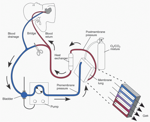
The typical ECMO circuit consists of tubing, a mechanical blood pump, membrane lung (oxygenator), cannulas for blood drainage (venous) and return (arterial), reservoir (bladder), heat exchanger, and pressure, flow, and oxygen saturation monitors (1,14). Figure 31.1 is an illustration of a typical ECMO circuit, with the circuit components discussed in detail later in the chapter. To provide cardiorespiratory support, blood is drained from the venous circulation into the ECMO circuit. A pump then propels blood through the membrane oxygenator for gas exchange. The oxygenated blood is warmed to the desired body temperature and returned to the patient via the arterial cannula.

ECMO is used in two distinct support modes: venoarterial ECMO (VA ECMO) and veno-venous ECMO (VV ECMO) (1,2,15,16). The differences between the two modes are listed in Table 31.1 and discussed below in detail.


Veno-Arterial ECMO

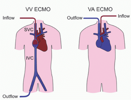
In this mode, blood from the venous circulation is drained into the ECMO circuit, pumped through the oxygenator for gas exchange, and the oxygenated blood from the circuit is returned to the arterial circulation (1,2,15,16). In this configuration, ECMO provides both cardiac and respiratory support (Figs. 31.1, 31.2).

Preload to the heart is decreased during VA ECMO because blood is drained from the venous circulation into the ECMO circuit (17,18,19). Decreased preload reduces myocardial contractility, left ventricular stroke volume, and pulse pressure. When the venous circulation is completely drained into the ECMO circuit, nonpulsatile flow ensues. However, in myocardial disease, decreased myocardial contractility and loss of pulsatile flow can occur prior to complete venous drainage, as injured or diseased myocardium may require higher

end-diastolic pressure and volume for myocardial contractility. When myocardial injury or disease improves, myocardial contractility returns, left ventricular ejection increases, and pulsatile flow resumes. Because decreased preload reduces contractile function of the ventricle, status of myocardial preload should be taken into account when evaluating myocardial contractility during ECMO.


FIGURE 31.1. ECMO circuit in veno-arterial support mode.


TABLE 31.1. Differences between veno-arterial and veno-venous ECMO modes


VA ECMO


VV ECMO


Support type


Cardiac and pulmonary


Pulmonary only


Cardiac support


Partial or complete


None


Cannulation sites


Venous and arterial sites


Venous sites


Cardiac preload


Decreased


Unchanged


Cardiac afterload


Increased


Unchanged


Pulmonary blood flow


Decreased


Unchanged


Pulse pressure


May decrease


Unchanged


Coronary blood flow


LV ejection or ECMO


LV ejection


Recirculation


None


Common


VA ECMO, veno-arterial ECMO; VV ECMO, veno-venous ECMO; LV, left ventricle.


FIGURE 31.2. Veno-venous (VV) and veno-arterial (VA) ECMO. SVC, superior vena cava; IVC, inferior vena cava.

Afterload to the left ventricle (LV) is increased during ECMO support (17,20,21). Increased afterload may be due to systemic vasoconstriction from sympathetic activation and the stress response to critical illness, use of inotrope and vasoconstrictor infusions, and arterial flow from the ECMO circuit into the aorta. Increased afterload increases LV end-diastolic pressure (LV-EDP), LV wall stress, and left atrial (LA) pressure (22,23,24,25,26). The manifestation of LA hypertension during VA ECMO is severe pulmonary edema, pulmonary hemorrhage, and “white-out” of the lung fields on chest X-ray (Fig. 31.3). Furthermore, increased LV-EDP and wall stress can impair myocardial recovery. Because the LV is not directly drained during VA ECMO, left heart decompression may be required in some patients supported with VA ECMO to lower LA pressure and reduce pulmonary edema and hemorrhage. Left heart decompression may also reduce LV wall stress and thereby promote myocardial recovery. Left heart decompression can be achieved in the interventional cardiac catheterization laboratory by creating an atrial communication (balloon atrial septostomy), or by draining the LA into the venous limb of the ECMO circuit using a drainage cannula (LA venting cannula).

Cardiac output during VA ECMO is a combination of the ECMO flow and native cardiac ejection. ECMO flow and systemic vascular resistance (SVR) determine mean arterial blood pressure (MAP) (18). In VA ECMO without native LV ejection, oxygenated blood from ECMO arterial return flows retrograde in the ascending aorta and aortic arch to provide coronary blood flow. However, in those patients with native LV ejection, coronary blood flow is antegrade from native LV ejection. Native LV ejection contains blood that is not drained into the ECMO circuit from the right atrium (RA) but ejected into the pulmonary circulation by the right ventricle and returned to the left heart. Mechanical ventilation with appropriate inspired oxygen concentration (Fio2) is required to oxygenate the blood transiting the pulmonary circulation and returning to the left heart in order to deliver oxygen to the myocardium. In an animal model of VA ECMO, Shen et al. (27) have shown that myocardial perfusion with oxygenated blood, provided by mechanical ventilation with an appropriate Fio2, was associated with improved myocardial recovery.


FIGURE 31.3. Pulmonary edema due to left atrial hypertension during veno-arterial ECMO. A: Pulmonary edema soon after ECMO cannulation; B: Resolution of pulmonary edema after balloon atrial septostomy.


Veno-Venous ECMO

In VV ECMO, venous blood is drained from the venous circulation into the ECMO circuit, pumped through the oxygenator, and returned to the RA (Fig. 31.2) (1,15,16,28). Oxygenated blood returned to the RA from the ECMO circuit enters the RV through the tricuspid valve (TV) and is ejected into the pulmonary circulation (Fig. 31.4). The oxygenated blood transits the pulmonary circulation to the left heart and is ejected by the LV into the systemic circulation. Thus, VV ECMO does not provide cardiac support and depends on native cardiac function to maintain cardiac output.

Systemic oxygen saturation (Sao2) depends on the proportion of oxygenated blood entering the right ventricle (RV)
(28,29). Therefore, positioning the return limb of the venous cannula such that oxygenated blood from the ECMO circuit returning to the RA is directed toward the TV is crucial (Fig. 31.4). Recirculation of some oxygenated blood returned to the RA back into the venous drainage is common. However, recirculation of significant amounts of oxygenated blood results in reduced Sao2. Recirculation can be assessed by comparing the partial pressure of oxygen in the arterial (Pao2) and venous (Pvo2) limbs of the VV ECMO circuit. A Pvo2 <20% of the Pao2 is acceptable and does not impair the efficiency of VV ECMO (30). Decreased Sao2 during VV ECMO should prompt the evaluation of cannula position to ensure that oxygenated blood returned to the RA is directed toward the TV. Other causes of low Sao2 during VV ECMO include hypovolemia, RV dysfunction, increased pulmonary vascular resistance (PVR), and developing oxygenator failure. These issues should be promptly investigated and treated.


FIGURE 31.4. Cannula position for veno-venous ECMO. SVC, superior vena cava; IVC, inferior vena cava; TV, tricuspid valve; RV, right ventricle; LV, left ventricle; Ao, aorta; PA, pulmonary artery.


Choosing an ECMO Mode

Patients with severe cardiac dysfunction due either to primary cardiac disease or secondary to other diseases (e.g., sepsis) require VA ECMO (1,14). VV ECMO is used to support children with respiratory failure with adequate cardiac function and output. Children with heart disease presenting with pure respiratory failure can be successfully managed with VV ECMO (31,32). Some patients with hypoxemic respiratory failure may have cardiac dysfunction secondary to hypoxemia. Restoration of systemic oxygenation with VV ECMO in these patients can improve cardiac function by correcting respiratory acidosis, reducing PVR, and enhancing myocardial oxygenation. In these patients, an initial trial of VV ECMO can help avoid the need for VA ECMO.

INDICATIONS FOR ECMO

Cardiac or respiratory failure unresponsive to conventional medical therapies is the usual indication for ECMO (Table 31.2) (1,14,33). ECMO has been used successfully to support neonatal respiratory failure due to persistent pulmonary hypertension and meconium aspiration syndrome. In children, respiratory failure due to pulmonary infection (bacterial, viral, or other pathogens) is a common indication for ECMO support. Cardiac indications for ECMO support include management of refractory cardiac dysfunction following cardiac surgery for congenital heart disease, and nonsurgical cardiac dysfunction due to myocarditis or cardiomyopathy (33). The availability of inhaled nitric oxide and high-frequency ventilation has reduced the need for ECMO in neonates with respiratory failure. Recent trends
indicate that ECMO is being increasingly used to support neonates and children with postoperative cardiac failure following surgery for congenital heart disease (34).


TABLE 31.2. Indications for ECMO


Indications


1.


Refractory hypoxemic or hypercapnic respiratory failure


2.


Refractory cardiogenic shock


Acute myocarditis


Cardiomyopathy


Severe sepsis


3.


Postoperative refractory cardiac failure


Postoperative low cardiac output syndrome


Failure to wean from cardiopulmonary bypass


Refractory arrhythmias


Pulmonary hypertension


4.


Cardiac arrest refractory to conventional cardiopulmonary resuscitation


5.


Procedural support


6.


Bridge to lung or heart transplantation or ventricular assist device


Relative contraindications


1.


End-stage primary disease with poor prognosis


2.


Severe neurologic injury or intracranial bleeding


3.


Uncontrolled visceral bleeding


4.


Prematurity (<34 wk gestation)


5.


Low weight (<2 kg)


6.


Family or patient directive limiting ECMO use


ECMO is being increasingly used to support children with cardiac arrest failing to respond to conventional cardiopulmonary resuscitation (ECPR) (35,36). In these patients, ECMO provides circulatory and respiratory support while the etiology for cardiac arrest is being investigated and treated. ECMO can also be used to support cardiac and respiratory function when undertaking surgical and cardiac catheterization procedures in critically ill patients whose risk of cardiac arrest while undergoing the procedure is deemed to be high (37,38).

When considering ECMO support, it is important to note that ECMO is only a support modality and does not treat the primary disease causing cardio-respiratory dysfunction. Thus, ECMO is most useful for those patients whose primary disease is reversible or can be successfully treated and associated with good prognosis. ECMO is generally considered short-term support (days to ˜2 weeks) as a bridge to recovery. However, some patients supported with ECMO for cardiac or respiratory failure fail to recover and wean off ECMO. In these patients, ECMO can be used as a bridge to heart or lung transplantation if the patient is deemed to be suitable for transplantation (39,40). The use of ECMO as a bridge to cardiac transplantation has been largely replaced with ventricular assist devices (VAD) as these devices provide mechanical support for longer duration (41,42). In some patients presenting with cardiogenic shock, ECMO can be used initially to resuscitate patients from shock prior to VAD implantation (bridge to VAD). ECMO remains an important mechanical support modality to bridge patients to lung transplantation.

Contraindications for ECMO support are relative and vary widely between institutions (Table 31.2) (33). ECMO is not useful when prognosis for the primary condition causing cardiac or respiratory failure is poor (irreversible or inoperable) or end-stage. Critically ill children with advanced multi-organ failure and those with severe neurologic injury may not benefit from ECMO. Anticoagulation used for ECMO support can exacerbate preexisting intracranial bleeding and bleeding in major organs, and thus ECMO may be deleterious for these situations. The risk of neurologic injury and intracranial hemorrhage is high in premature infants (<34 weeks gestation). Small-sized neonates (<2 kg) may pose technical challenges to ECMO cannulation and support. Finally, patient and family directives may limit the use of ECMO support in some patients.


EQUIPMENT FOR ECMO


Circuit Tubing and Prime

ECMO circuit tubing is made from a polyvinylchloride-based plastic compound (43). Blood contact with the prosthetic surface of the ECMO circuit tubing activates the coagulation cascade and platelets to form clot (44). The inflammatory cascade is also activated, resulting in a systemic inflammatory response and capillary leak syndrome. Biocompatible circuit-lining materials (e.g., Carmeda BioActive Surface [CBAS] uses heparin coating; Carmeda, Stockholm, Sweden) that reduce blood contact activation have been used in ECMO circuits to reduce clotting (45,46). Release of plasticizer (di-2-ethylhexyl phthalate [DEHP]) has been a concern with the use of polyvinyl tubing (47). Exposure to DEHP has shown to be associated with infertility, reduced sperm production, and ovarian dysfunction in animal studies (48). However, a study of adolescents exposed to DEHP during neonatal ECMO support by Rais-Bahrami et al. (47,49) showed no effects on physical growth and pubertal maturity.

ECMO circuits can be primed with either blood or crystalloid (e.g., Plasma-Lyte, Abbott Laboratories, Abbott Park, IL) solution. Blood-primed ECMO circuits are generally used when ECMO is deployed semi-electively, whereas crystalloid-primed circuits are commonly used for urgent ECMO support such as in patients cannulated for ECMO during cardiopulmonary resuscitation (CPR) (33,36).


Blood Pump

The function of the blood pump is to propel blood through the oxygenator and ECMO circuit (43,50). Ideally, an ECMO pump should be able to provide a wide range of flows (75-200 mL/kg/min) without exerting excessive shear stress on red blood cells and causing hemolysis. Currently available ECMO pump technology can be categorized into two major types: roller and centrifugal. Comparisons between the two pump types are listed in Table 31.3.

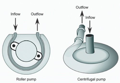
Roller pumps are positive displacement pumps that use a rotating roller head to sequentially compress linear tubing against a back plate, thereby forcing the column of blood in the tubing forward (Fig. 31.5) (43,50). The portion of the ECMO tubing within the pump is called the “raceway” and is
made of reinforced material (e.g., Tygon S-95-E; Tygon, Saint-Gobain Corp., Courbevoie, France) to help withstand the shear stress imposed by the roller heads (51). Pump output is a function of pump speed (revolutions per minute; RPM), raceway tubing diameter, and degree of occlusion (43,50). To avoid direct suction to the venous catheter in roller pump ECMO circuits, a reservoir called the “bladder” is placed between the pump and the venous drainage tubing (52). Venous blood from the patient is drained passively by gravity into the bladder, limiting the direct application of negative suction pressure to the venous inflow and preventing injury to vascular structures and endothelium. A servo-control mechanism contained in the bladder regulates pump flow by decreasing pump speed when the bladder has reduced volume or is empty. This servo-control mechanism prevents cavitation and hemolysis from excessive negative pressure resulting from continued pump function when pump venous inflow is reduced or occluded and is thus an important safety feature for roller-pump ECMO circuits (53).


TABLE 31.3. Comparison of roller and centrifugal pumps


Roller pump


Centrifugal pump


Pump mechanism


Positive displacement


Centrifugal force


Pump inflow


Passive drainage


Active drainage


Pump occlusion


Occlusive


Nonocclusive


Retrograde flow


Not possible


Possible


Pump flow


More precise


More variable


Factors affecting pump flow


Pump RPM Displacement volume


Pump RPM, Preload and afterload


Risk of tubing rupture with outflow obstruction


Present


Extremely rare


Hemolysis


Present


Present


RPM, revolutions per minute.


FIGURE 31.5. Schematic diagram of roller and centrifugal pumps.

Pump outlet pressure is a function of pump speed and resistance in the tubing, oxygenator, cannula, and SVR (43,50). In roller pumps, when the ECMO circuit (i.e., tubing, oxygenator, or arterial cannula) beyond the pump outlet is obstructed, the pump will continue to flow resulting in high outlet pressure proximal to the obstruction which can result in circuit tubing rupture. Thus, a high outlet pressure alarm is essential and should prompt careful evaluation for ECMO circuit outflow obstruction.

Centrifugal pumps contain a spinning rotor in a rigid conical housing that creates a constrained vortex to draw blood into the pump and uses centrifugal force to eject blood from the pump (Fig. 31.5) (43,50). Sub-atmospheric pressure is created at the center of the vortex and pump inlet, resulting in active drainage of blood from the patient into the pump. Positive pressure created at the edge of the vortex results in passive ejection of blood through the pump outlet creating pump flow. Centrifugal pumps are nonocclusive systems and create continuous nonpulsatile flow. The centrifugal pump rotary mechanism will continue to spin when venous inflow becomes obstructed resulting in increased negative pump inlet pressure causing hemolysis and rarely cavitation. These issues can be overcome with reducing pump speed at times of decreased venous return or incorporating a bladder to automatically regulate pump speed based on pump inflow. Because ejection of blood from the centrifugal pump is passive, pump flow for a given pump speed is dependent on resistance applied to pump outflow from the ECMO circuit and SVR. Increased outflow resistance can decrease pump flow. Circuit rupture with outflow occlusion is extremely rare because pump flow is reduced or absent with increased resistance. Because centrifugal pumps are nonocclusive systems, retrograde flow from the outlet into the pump is possible at low pump rotational speeds.

Other potential problems with centrifugal pumps relate to issues of blood stagnation and heat generation in the pump (43,50). These issues can result in thrombus formation within the pump and hemolysis. Newer generation centrifugal pump designs have reduced stagnation, heat generation, and shear stress to blood cells and have helped minimize these complications. Even though centrifugal pumps are becoming increasingly used in ECMO circuits, reports comparing complications and patient outcomes with roller pumps have shown mixed results. Some studies have shown increased hemolysis with roller pumps, and others show no difference in pump-related complications and patient outcomes (54,55

Only gold members can continue reading. Log In or Register to continue

Stay updated, free articles. Join our Telegram channel

Jun 7, 2016 | Posted by in RESPIRATORY | Comments Off on Extracorporeal Membrane Oxygenation in Infants and Children

Full access? Get Clinical Tree

Get Clinical Tree app for offline access