Ex Vivo Arterial Repair for Renovascular Hypertension Secondary to Fibrodysplasia


Ex Vivo Arterial Repair for Renovascular Hypertension Secondary to Fibrodysplasia


Michel J.T. Visser and J. Hajo van Bockel


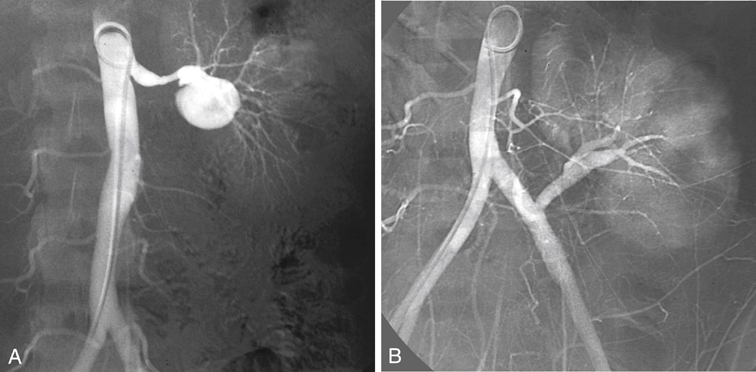
Fibrodysplastic disease of the renal arteries is bilateral in more than half of the patients, and involvement of the primary branches of the renal artery has been reported in as many as 40%. In most patients the fibrodysplastic lesions are located in the distal renal artery, but many lesions affect multiple smaller renal arteries or extend into the primary and secondary branches of the renal artery. In these latter patients, percutaneous angioplasty is not without complications, and dissection, occlusion, renal infarction, perforation, and contrast-induced kidney failure have been reported (Figure 1). Open reconstructions are favored in many of these patients, yet prospective data comparing the results of transluminal angioplasty and open surgical repair are available for treatment of atherosclerotic lesions but not for fibrodysplasia.


Therapy


Surgical reconstruction is considered the gold standard of therapy for renal fibrodysplastic lesions. Operative morbidity and mortality rates in these young patients are very low, and excellent long-term functional results up to 15 years or more have been reported by many. A variety of surgical techniques have been used for surgical revascularization for repair of stenosis in the main renal artery. Lesions extending into the renal arterial branches can be repaired by both in situ and ex vivo techniques. In these circumstances, we prefer extracorporeal reconstruction and autotransplantation of the kidney.


The advantages of ex vivo reconstruction are optimal exposure, a bloodless surgical field, protection of the kidney from prolonged warm ischemia, no time limit for microvascular repair, and contraction of the kidney resulting from preservation, which allows an easier dissection. Finally, the complete reconstruction can be tested for both patency and leakage before implantation. Compared with in vivo repair, ex vivo repair is associated with longer operation times, more blood loss, and more wound hematomas. Although it has been suggested that stenosis at the renal bifurcation can be repaired in situ, results of extracorporeal repair appear to be superior. Standard indications for performing ex vivo arterial repair exist (Box 1).


Surgical Technique


Ex vivo procedures consists of three parts: removal and preservation of the kidney, ex vivo reconstruction, and autotransplantation of the kidney into the patient. The technique has been adapted from kidney transplantation and was introduced by Gelin and Belzer early in the 1970s.


Incision


The incision depends on the personal preference and experience of the surgeon. Ex vivo reconstruction can be performed through either a midline or retroperitoneal incision. The midline incision is used when a bilateral procedure is performed simultaneously. If a unilateral reconstruction is planned or a bilateral procedure is staged, a retroperitoneal incision is preferred.


The patient is positioned prone or, alternatively, the flank may be slightly elevated with a beanbag. The retroperitoneal incision preferred is slightly different from the standard incision as used by others. It usually extends from the costal arch (the incision is often extended between the ninth and tenth or the eighth and ninth ribs if more exposure is required in obese patients) and slightly medially curved just cranial to the pubic bone. In male patients the lateral rectus abdominis muscle may be partially transected. The retroperitoneal space is entered in the proximal part of the wound to avoid opening of the peritoneum.


By blunt dissection, the peritoneum and its contents can be swept medially. A fixed retractor (Omnitract) greatly facilitates the nephrectomy, the mobilization of the ureter, and, if required, the harvest of a branched hypogastric autograft through the one incision. Intravenous fluids are liberally infused to ensure adequate hydration and to promote renal vasodilation and optimal urine production.


Nephrectomy


The objective of the nephrectomy part of the procedure is to remove the kidney without damage to the arterial tree and to optimally preserve its function. The nephrectomy is not much different from the procedure in a living kidney donor and may even be performed laparoscopically.


The kidney involved is dissected, avoiding unnecessary manipulation and traction to prevent renal vasospasm, which interferes with its preservation. Gerota’s fascia is opened at the level of the lower pole. Subsequently, the anterior and posterior part of the kidney is dissected sharply. This can be difficult if a partial infarction is present and the perinephric fat is densely adherent to the kidney. The hilar part of the renal artery is not dissected, and it is preferred not to enter the hilar tissue between the upper and lower pole of the kidney and the aorta (left-hand side) or the caval vein (right-hand side) to prevent accidental injury or spasm of small branches. If early branching of the right renal artery is present, the inferior vena cava may be mobilized, which involves suture ligation and transection of a pair of lumbar veins at the level of the renal vein. Significant collateral circulation is interrupted only after the dissection has been completed.


The kidney is then left alone to recover from possible vasospasm. The ureter is dissected free down to the entrance into the bladder, taking care not to disturb its blood supply. As soon as hydration is adequate, mannitol is infused around 10 minutes before arterial occlusion to promote renal perfusion and a diuresis, lessening the risk of acute tubular necrosis.


The patient is anticoagulated with heparin sodium. The ureter is clamped with a supple tourniquet (vessel loop) during the cold ischemic period of preservation and extracorporeal repair. This maneuver prevents, on the one hand, warming of the kidney by collateral ureteral circulation toward the kidney and, on the other hand, leakage of potassium-rich preservation fluid in the patient’s circulation. Subsequently, the artery is occluded with a right-angle clamp close to the aorta, and a Satinsky clamp is placed on the renal or the caval vein, which allows a small venous patch to be taken from the cava.


The kidney is removed and placed on melting ice-slush in a flat basin on top of the patient’s pubic bone. The artery and, occasionally, large collaterals are cannulated with small cannulas and flushed with approximately 300 mL of extracellular renal preservation solution at 4°C until the venous effluent becomes clear and the entire kidney is bloodless and pale. For each branch, a separate cannula and a separate line are used. No ligatures are applied. Leakage between the catheter and vessel is easily prevented by digital pressure only.


The preservation fluid drains only by gravity; pressure-increasing devices are never required and can damage the kidney. If the renal artery or branches are occluded, as can occur in renal artery dissection, these branches are dissected free, incised until the lumen is opened, and cannulated using separate lines. Care is taken to avoid rotating the kidney during the explantation in order to avoid twisting of the ureter during autotransplantation. Although others have recommended machine preservation in the past, this practice has been abandoned. It makes the operation more complicated and has no benefit. Subsequently, the kidney is placed in a small basin on the top of the pubic bone. The kidney is covered with melting ice for continuous hypothermia, leaving the hilar structures for dissection.

Stay updated, free articles. Join our Telegram channel

Aug 25, 2016 | Posted by in CARDIOLOGY | Comments Off on Ex Vivo Arterial Repair for Renovascular Hypertension Secondary to Fibrodysplasia

Full access? Get Clinical Tree

Get Clinical Tree app for offline access