>20–40 mm
>40 mm
0–30 mm
>30 mm
18
5
43
11
66.6%
100%
72.1%
81.8%
0.378
>10–20 mm
>20–30 mm
>30 mm
16
7
13
43.7%
71.4%
76.9%
>20 mm
67
89.6%
15–20 mm
>20 mm
14
71
71%
87%
ENB = Electromagnetic navigation bronchoscopy, FDG-PET = fluorodeoxyglucose positron emission tomography, ROSE = rapid on-site examination of cytopathology.
High-risk nodules (>60% risk of malignancy)
Nodules that have radiographic features that are high risk for malignancy should be managed operatively with resection in patients who can tolerate surgery. Preoperative workup should consist of a thorough evaluation of fitness for surgery, including pulmonary function tests and assessment of baseline functional status. Surgery is performed via video-assisted thoracoscopic surgery (VATS) or robotic techniques whenever possible. We rarely recommend that a thoracotomy be performed. If a diagnosis has not been established, a wedge resection with adequate (R0) margins should be performed with frozen histologic assessment. Benign lesions, typical carcinoid tumors, and metastatic lesions from other sources (i.e. sarcoma, renal cell carcinoma) require no further resection. Currently, the standard of care for non-small-cell lung cancer in patients with adequate pulmonary function tests (PFTs) is anatomic lobectomy with mediastinal lymph node sampling or dissection. It is worth noting that small lesions, particularly GGOs and semisolid nodules, may be difficult to locate by manual palpation, particularly using VATS techniques. In these patients, we have found that radiotracer-guided resection with technetium-99m microalbumin aggregate safely facilitates resection of the nodule and is cost-effective[34–36].
Conclusions
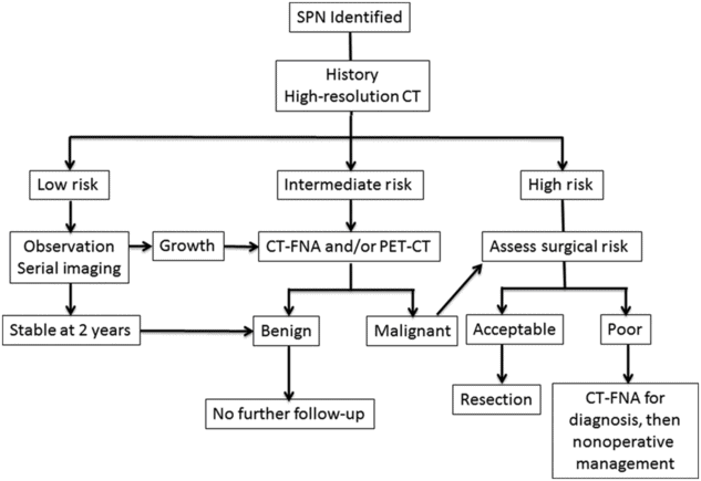
SPNs are becoming increasingly common clinical entities, and thus thoracic surgeons should be comfortable with the evaluation and management of these lesions (Figure 11.1). Patients can be risk stratified into low-, intermediate-, and high-risk groups for malignancy based on a thorough history and review of radiographic studies. High-resolution CT scan is an important component of the initial workup. In general, low-risk nodules should be followed with serial imaging, intermediate-risk nodules should trigger additional workup via CT-FNA and/or PET-CT, and high-risk nodules should be managed surgically in patients with acceptable operative risk.

Workup algorithm for SPNs.
References
Stay updated, free articles. Join our Telegram channel

Full access? Get Clinical Tree


