Esophageal Perforation
Presentation
A 44-year-old man presents to the emergency department after a night of binge drinking complaining of dysphagia and odynophagia. He admits to having vomited several times. Physical findings include tachycardia, fever (temperature 100.8°F), and decreased breath sounds at the left base.
Differential Diagnosis
In a patient who presents with esophageal symptoms after episodes of vomiting, esophageal perforation must be strongly considered. Other possible diagnoses include aspiration pneumonia, esophagitis, and peptic ulcer disease.
Discussion
Barogenic esophageal perforation due to emesis is a well-recognized cause of esophageal perforation. It typically occurs in younger men and is often related to an episode of alcohol abuse. The rupture usually occurs in the distal esophagus, permitting saliva and gastric contents to leak into the mediastinum and possibly the pleural spaces. The peristaltic activity of the esophagus and negative intrathoracic pressure facilitate rapid accumulation of these digestive fluids, leading to inflammation and the development of multisystem organ failure if the condition is left untreated.
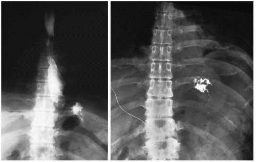
A high index of suspicion is appropriate when esophageal perforation is part of the differential diagnosis. A contrast esophagram is useful and should be performed in patients who are able to cooperate with the procedure. Radiography permits accurate assessment of the site of the leakage and the cavity into which the leakage drains. In patients who are not able to cooperate with such an examination, upper endoscopy is performed.
The site of the leakage helps determine how to approach the patient surgically. Patients who have mid-esophageal leaks are best approached through a right thoracotomy because the aortic arch interferes with adequate access from the left chest. Most other patients are approached through a left thoracotomy because the lower esophagus is more readily accessible and because the left pleural space is more often involved in the process.
Recommendation
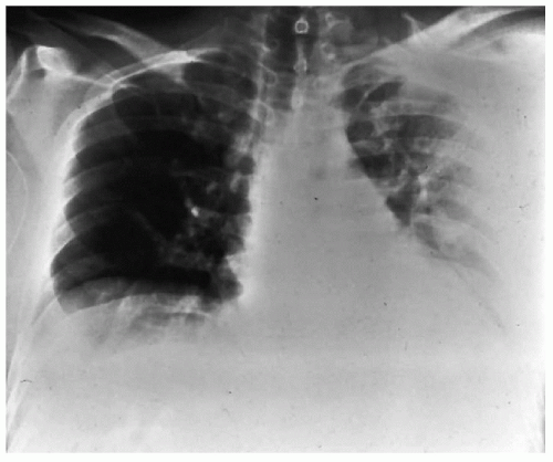
Chest x-ray. Contrast esophagram.
▪ Chest X-ray
Chest X-ray Report
Small left pneumothorax, large left pleural effusion. ▪
▪ Contrast Esophagram
 Figure 25-2
Stay updated, free articles. Join our Telegram channel
Full access? Get Clinical Tree
 Get Clinical Tree app for offline access

|
