Enlarged Cardiac Silhouette
Gregory Kicska, MD, PhD
DIFFERENTIAL DIAGNOSIS
Common
Ischemic Cardiomyopathy
Valvular Disease
Heart Failure Exacerbation
Pericardial Effusion
Less Common
Nonischemic Dilated Cardiomyopathy
Pericardial Mass
Rare but Important
Left Ventricle Aneurysm
ESSENTIAL INFORMATION
Key Differential Diagnosis Issues
Pericardial space fluid: Globular enlargement
Cardiac chamber enlargement: Characteristic contour abnormality such as filling of retrosternal clear space in right ventricle enlargement
Pericardial mass: Focal contour irregularity
Helpful Clues for Common Diagnoses
Ischemic Cardiomyopathy
Sub-endocardial fat or calcium, left ventricle (LV) wall thinning in coronary distribution, dense coronary calcifications
MR shows subendocardial or transmural delayed enhancement in coronary artery distribution
Valvular Disease
Valvular calcifications most common
MR cine or phase contrast shows flow jets
Heart Failure Exacerbation
Coexistent signs of pulmonary edema
Pericardial Effusion
New globular heart enlargement on radiograph, fluid-density pericardial fluid on CT
Hemopericardium suggested by high-density pericardial fluid or neoplasm history (lung, breast, melanoma)
Helpful Clues for Less Common Diagnoses
Nonischemic Dilated Cardiomyopathy
Dilated LV, thin wall, EF < 40%
Either no delayed enhancement present or enhancement is not subendocardial
Pericardial Mass
Pericardial cyst: Circumscribed fluid density at right more than left cardiophrenic angle
Pericardial fat pad: Fat density most commonly at right cardiophrenic angle
Helpful Clues for Rare Diagnoses
Left Ventricle Aneurysm
True aneurysm
Post infarct wall thinning, dilatation, and associated thrombus
Most often present along apical anterior or lateral wall
False aneurysm
Ruptured myocardium contained by pericardial adhesions at inferior-basal wall
Neck narrower than internal diameter
Image Gallery
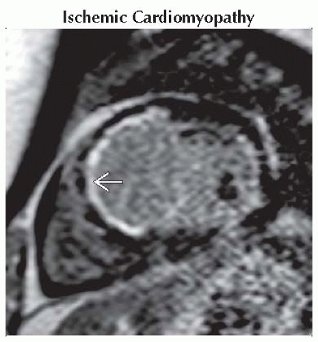 Short axis delayed gadolinium-enhanced image shows subendocardial enhancement
 in the septal and anterior wall at the base. The patient had hypokinesis and wall thinning at this location. in the septal and anterior wall at the base. The patient had hypokinesis and wall thinning at this location.Stay updated, free articles. Join our Telegram channel
Full access? Get Clinical Tree
 Get Clinical Tree app for offline access

|