Endovascular Treatment of Aortic Dissection


Endovascular Treatment of Aortic Dissection


David M. Williams and Himanshu J. Patel


Acute aortic dissection causes death by rupture of the false lumen or by end-organ malperfusion. Untreated acute type A dissection has a mortality of approximately 50% within 48 hours, predominantly caused by rupture resulting in cardiac tamponade. Acute type B dissections have a mortality of approximately 25%, which doubles when the dissection is complicated by paraplegia or by renal, mesenteric, or lower extremity ischemia.


In recent years, numerous publications have described endovascular techniques for addressing the complications of acute type B and selective type A dissections, and as well as for elective and emergent procedures to treat chronic dissection. Endovascular techniques for treating aortic dissection fall into two classes: preventing rupture by excluding the false lumen by means of a carefully deployed endograft and treating malperfusion by correcting branch artery obstruction by means of an endograft or fenestration, supplemented by branch artery stents.


Role of Imaging


Despite the enthusiasm for endovascular treatment of aortic dissection, standard open repair of the uncomplicated type A dissection will remain the predominant life-saving procedure in this patient cohort for at least the next 5 to 10 years. Clinical examination and expeditious imaging of patients with transesophageal ultrasound or CT are the mainstay of early diagnosis of aortic dissection and of the triage of patients between emergent open repair versus medical or endovascular management.


High-quality cross-sectional imaging is critical for planning and performing endovascular procedures for aortic dissection. Features crucial for planning include evidence of contrast extravasation or contained rupture, location of large intimal tears, relation of tears to critical aortic branches, the identity and anatomic relation of the true and false lumens, the longitudinal extent of the dissection, the distribution of critical branches from the true and false lumen, the relation of the dissection flap to critical branch origins, and evidence of malperfusion.


Endovascular interventions can result in dramatic changes in the aortic anatomy and branch vessel perfusion. These changes include extension of dissection from type B to type A, tearing and intussusception of the aortic intimal flap, and collapse of the true lumen with new obstruction of branch arteries. Successful management of such complications requires prompt recognition of the presence and extent of the anatomic change, which requires intraoperative imaging by intravascular ultrasound (IVUS), transesophageal echocardiography (TEE), and angiography. IVUS provides the best mix of broad anatomic coverage, detection of changes in dissection flap configuration, and precise anatomic demonstration of the relation of the flap and vessel origins. Angiography allows assessment of branch artery perfusion flow and pressure. TEE is often used as an intraoperative monitor and gives information about changes in thoracic aortic anatomy similar to that from IVUS.


Vascular Access


Endovascular treatment of an aortic dissection usually entails at least two access sites. Imaging is critically necessary for constant orientation indicating in which lumen each guidewire is located. Because straight or angled guidewires can easily and inadvertently pass through intimal tears or small holes in the dissection flap from abrupted intercostal or lumbar arteries, one must always verify a guidewire’s location before deploying aortic stents or endografts. Most of these orientation checks are easily accomplished by IVUS, which some practitioners consider an essential tool in treating dissections.


Access for placement of an endograft currently requires one femoral artery cutdown and one percutaneous access. Access for fenestration requires access through two femoral arteries, usually entailing one access in each groin, occasionally tandem accesses in a single groin. Great effort should be made to enter the vessel by way of the true lumen, which simplifies treatment of many variants of branch artery malperfusion. Although particular interventions such as placement of an aortic stent or stent graft require absolute certainty of guidewire location in the true lumen throughout the treated segment, monitoring of procedural activities by IVUS can proceed from within either lumen.


Excluding the False Lumen


The premise of endograft treatment of aortic dissection is that exclusion of the intimal tear isolates the false lumen locally from perfusion pressure and turbulence of blood flow across the flap, and it restores preferential perfusion to the true lumen of the aorta distal to the intimal tear. Initial experience bears out the short-term success of this treatment of acute dissection, with local thrombosis on the false lumen adjacent to the endograft, elimination of dynamic obstruction of branch vessels, and variable relief of static obstruction. The size, number, and location of distal intimal tears affect the comprehensiveness of this hemodynamic benefit. Trials of endograft treatment of uncomplicated acute type B dissections are under way outside the United States (including ADSORB [Acute Dissection treatment with Stent graft OR Best medical therapy] supported by W. L. Gore and Associates, Flagstaff, AZ).


The dissected aorta presents unique challenges to endograft technology. In acute dissections the false lumen dilates and the true lumen collapses. In the classic (double-barrel) aortic dissection, the total aortic diameter is capable of acutely expanding to 20% of its baseline size. After exclusion of the acute entry tear by an endograft, the false lumen can collapse as the endograft expands. In the setting of an intramural hematoma, when the false lumen is largely thrombosed and not capable of immediate contraction, the endograft continues to exert force, especially at its upper and lower edges, against the delicate intimal flap overlying the thrombus. Treatment planning entails selecting an endograft that will have long-term stability and safety, being neither so small that it will migrate after the true lumen reexpands and the false lumen contracts, nor so large that it will tear the intimal flap by constant expansion against a thrombus-filled false lumen.


Malperfusion


Both endograft placement and fenestration with stenting are undertaken to treat obstruction of branch arteries. Although the diagnosis of malperfusion is a clinical decision, the premise of endovascular treatment is that ongoing arterial obstruction is maintaining the malperfusion and that relieving the obstruction will correct the malperfusion. Our own practice is to demonstrate that arterial obstruction is present by comparing pressures in the branch arteries to pressure in the aortic root.


In the absence of proof of ongoing obstruction, we do not perform fenestration or stenting or, if the false lumen is stable, deploy an endograft. An alternative approach to the clinical diagnosis of malperfusion is to assume without verification that it is caused by arterial obstruction, cover the entry tear with an endograft, and assess branch arteries after treatment. Although this reduces the duration of the procedure, forgoing baseline arterial assessments and failing to document ongoing arterial obstruction adds complexity to the task of evaluating the benefit of the procedure and uncertainty in trying to distinguish between an incomplete hemodynamic response versus a complication of endograft intervention.


Treatment of branch artery obstructions that are directly related to the dissection flap depends on whether the obstruction is static or dynamic. In a static or fixed obstruction, the stenosis is elastic and is caused by the dissection flap or hematoma extending into the branch artery. Treatment requires deployment of a stent across the stenosis. As noted, when the static obstruction is extensive, restoration of blood flow to multiple branches can result in collapse of the true lumen, with new dynamic obstruction of the branches of the true lumen. Intravascular ultrasound monitoring during the interventional procedure is a convenient method to monitor the behavior of the dissection without multiple contrast injections.


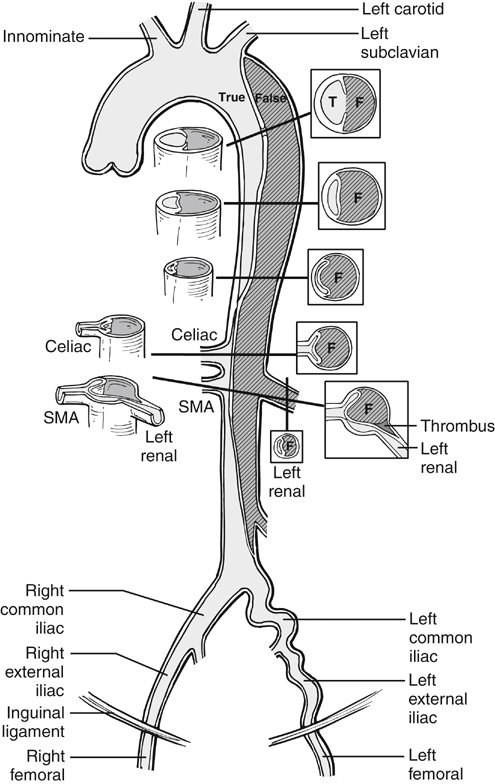
In dynamic obstruction, the true lumen is collapsed, and the flap spares the origin but prolapses across it. Generally several visceral branches are involved simultaneously. Treatment is reducing the flap and providing perfusion to the branches. This is most expeditiously accomplished by deploying an endograft, if the entry tear is anatomically suitable, as in many type B dissections. The endograft excludes the entry tear, diverts blood flow back exclusively into the true lumen, and simultaneously reexpands the true lumen and collapses the false lumen. Placement of an appropriate endograft results in global relief of distal true lumen collapse and dynamic obstruction, and it results in variable relief of static obstruction as well. Following placement of the endograft, perfusion of infradiaphragmatic branches can be assessed and, if indicated, treated by standard clinical and angiographic means (Figures 1 and 2). Three phase II trials of endograft treatment of complicated type B dissections are active in the United States at the time of this writing (W.L. Gore and Associates, Flagstaff, AZ; Medtronic, Minneapolis, MN; and Cook, Bloomington, IN).


If the aorta with collapsed true lumen is anatomically unsuitable for an endograft, such as when the tear is in the ascending aorta or the arch, then a fenestration procedure can be performed. Our own practice in cases of type A dissection depends on the duration and gravity of the malperfusion. Patients without prolonged malperfusion undergo immediate open repair of the ascending aorta. Patients with prolonged malperfusion, particularly of the legs, gut, or spinal cord, first undergo fenestration. If they survive the reperfusion injury, they subsequently have open repair of their aorta.


In the fenestration procedure, a tear in the dissection flap is created using intravascular ultrasound and fluoroscopic guidance, near the critical affected branch arteries. This is generally either near the level of the celiac trunk and just below the level of the renal arteries. A transjugular intrahepatic portosystemic shunt (TIPS) needle or other suitable curved needle is poised perpendicular to the flap and pushed through it, using a short, controlled jab (Figures 1 and 3). The hole in the flap is subsequently dilated with a 14-mm or similar angioplasty balloon, resulting in a transverse tear in the aorta, whose edges are drawn apart in a biconcave configuration by inherent longitudinal tension in the dissection flap (Figure 3D). Occasionally, intravascular ultrasound shows immediate relief of true lumen collapse. If collapse of the true lumen persists, a large-diameter self-expanding stent (such as 16 mm) is deployed in the aortic true lumen.


Stay updated, free articles. Join our Telegram channel

Aug 25, 2016 | Posted by in CARDIOLOGY | Comments Off on Endovascular Treatment of Aortic Dissection

Full access? Get Clinical Tree

Get Clinical Tree app for offline access